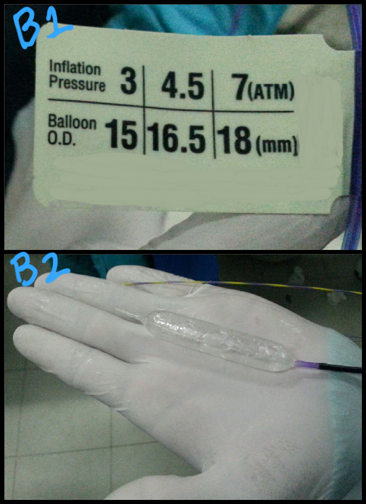
This 28 year old gentleman had what appeared to be "a soft mass with acoustic shadow" in his distal CBD. Contrast injection on ERCP showed a rounded stone in the mid CBD (A: green arrow). After a wide papillotomy, we attempted to extract the stone with an extraction balloon but were unsuccessful. We decided to do a sphincteroplasty with a wire guided balloon (B1 & B2: the balloon specifications and inflation check prior to insertion. C1 to C4: the balloon being placed into the papilla and inflated. C5: green arrows show the extent of dilated balloon on fluoroscopy). Even after sphincteroplasty, the stone could not be removed with the extraction balloon and we could clearly see it stuck in the CBD through the wide papillotomy (D: green arrow). We then decided to use the extraction basket to remove the lodged stone (E: white arrows show the open basket in the CBD). This was rewarded with success and the stone was removed (F: green arrow shows the stone, smeared with blood, lying in the duodenal lumen).


No comments:
Post a Comment