Saturday, April 26, 2014

Migrated biliary stent

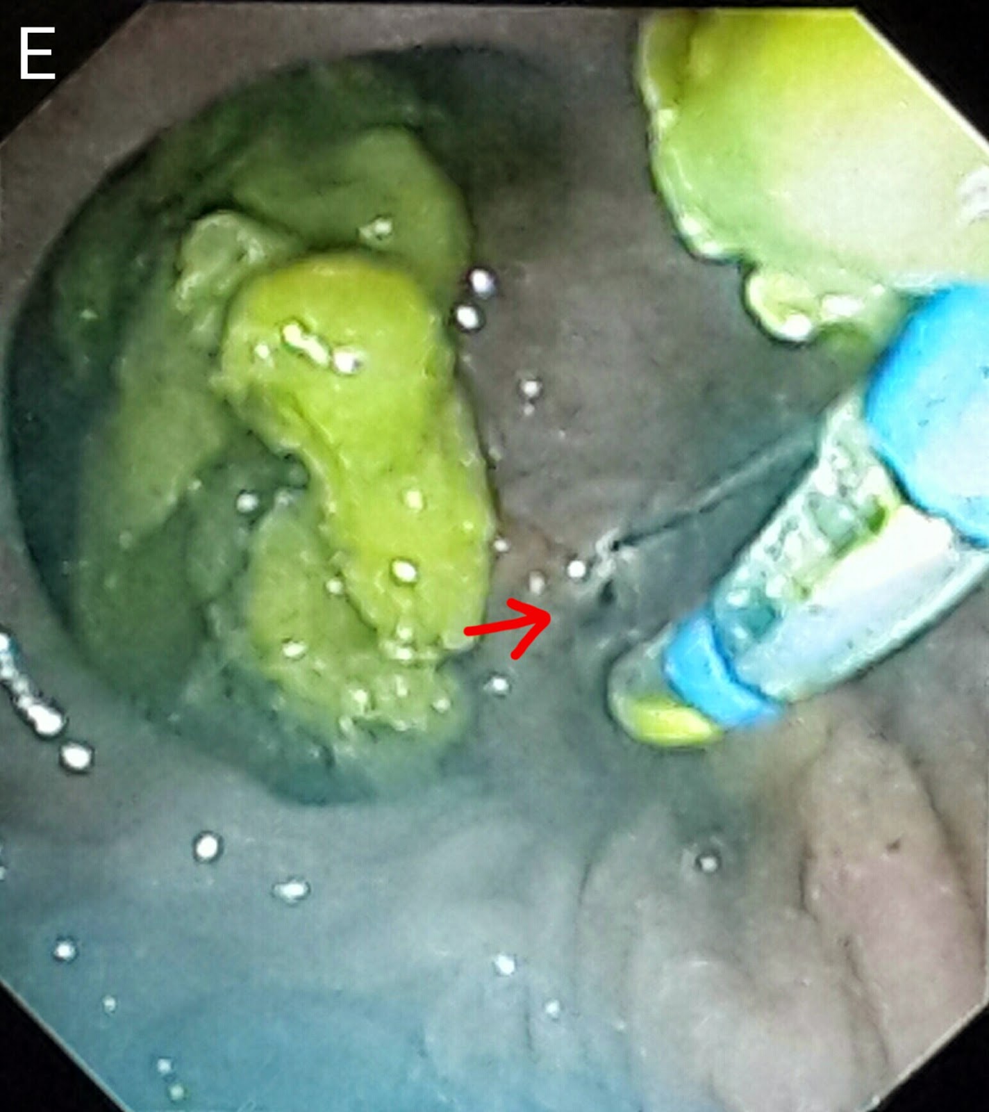
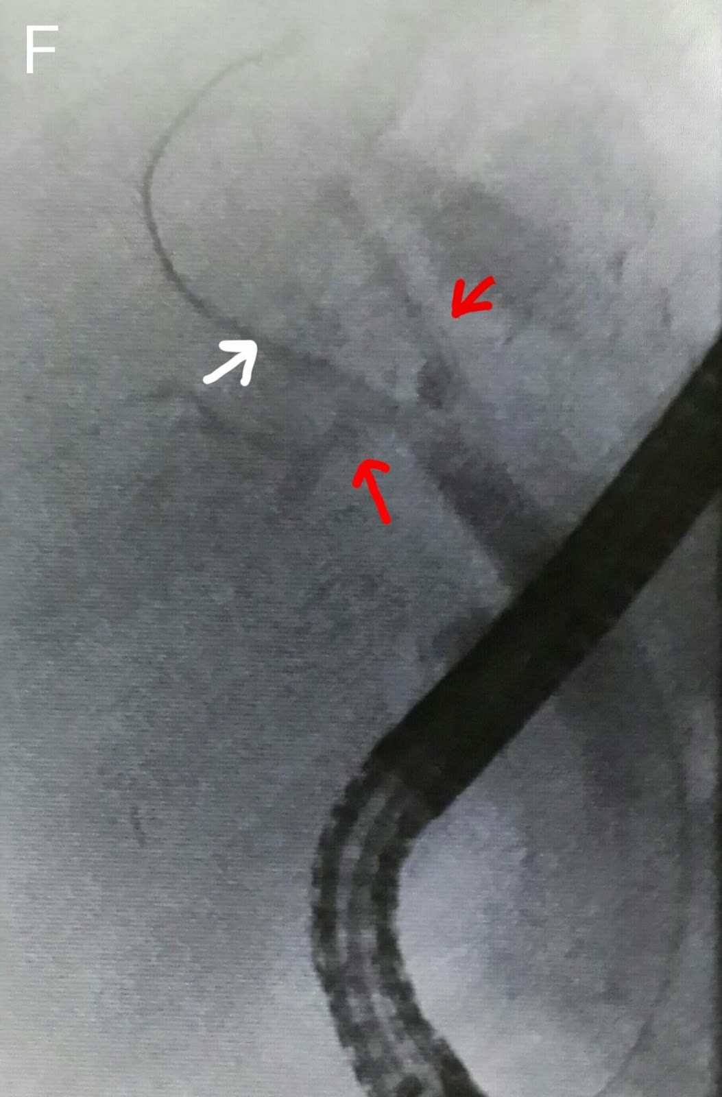
This 40 year old gentleman is an old patient of ours. He has been treated for hepatitis C and extra hepatic portal venous obstruction. He has been regularlyundergoing ERCPs at our center for bile duct obstruction. We usually clear the CBD and place a new plastic stent at each visit. This time around we couldn't see the previously placed stent and suspected that it had migrated into the bile duct. This was confirmed on flouroscopy (A: black arrows). We first attempted to pull the stent out using a stone extraction basket (B: white arrows show the stent. Black arrows mark the basket). This was unsuccessful so our next maneuver was to thread the stent with a guidewire (C: white arrows show the stent and black arrows mark the guidewire). The wire was passed to thread a Soehendra stent retriever over it (D: white arrow). The stent was properly engaged (E: white arrow indicates the stent retriever and black arrow market the engaged lower end of the stent). The old stent was then pulled out (F: black arrow shows the friable sludge ridden end of the stent being pulled by the Soehendra stent retriever-white arrow). We could have directly pulled free stent out through the scope channel but were concerned that it might break since the distal end was very cracked and friable. We used a snare to pull it out (G: white arrow shows the snare. H: The retrieved stent l. A wide papillotomy was done (I: white arrow). The CBD was swept with a stone extraction balloon (J: black arrow shows the inflated balloon in the bile duct). A lot of debris was removed (K: white arrows). Finally a 10 French diameter plastic stent of 12 cm length was placed (L: black arrows). This was a stent with a curved proximal end. Not the the classical pigtail shape. This would help in retention of the stent keeping in mind the papillotomy. 

Friday, April 25, 2014

Extra large diverticulae flanking ampulla

This 60 year old gentleman had been referred following the development of classical obstructive jaundice signs. His ultrasound reported an ampullary mass lesion. The CT report showed a greatly distended CBD with a large calculus at the terminal end (A: white arrow posts to the stone and black arrows delineate the CBD). There was a "whoa" moment the moment the papilla came into view as it was flanked by two huge diverticulae stuffed with food (B: red arrows. White arrow points to the papilla). The scope kept slipping and cannulating the ampulla was trickier than expected (C). Contrast injection showed a dilated CBD with a large stone near the lower end (D: black arrow). We did a small sphincterotomy (E: red arrow). A 10 French plastic stent of 10 cm length was then deployed (F1: red arrow shows the stent and blue arrow points to the stent assembly. F2: black arrows show the stent). The patient was referred to the surgeon for cholecystectomy and bile duct clearance. 

Lost to follow up

This 45 year old gentleman had suffered from bile duct injury whilst undergoing an open cholecystectomy 10 months ago. The surgeons placed a drain (A: red arrow shows the drain. White arrow shows the large leak-almost looking like the gallbladder in shape. Blue arrow shows the main bile duct). An MRI was done soon after and confirmed the leakage (B: red arrows show the bile leak). He was reffered to our facility and we had placed a 10 French 10cm long plastic stents to bridge the leak. (C: white arrows show the stent) . This was about ten months ago. He was then lost to follow up. He resurfaced at our outpatient out of curiosity asking what was to be done about the Stent we had placed. When questioned as to why he hadn't turned up a month after the procedure, he said he had been told by his physician that his condition has been cured and he needs no more treatment. The moment we went in for ERCP, we were greeted by a normal ampulla with no sign of his placed stent. We thought it had migrated inwards but that wasn't the case as it didn't show up on flouroscopy either. One thing we noted was almost absent bike flow from the ampulla and normal flow from another opening just above it (D: Blue arrow marks the ampulla and white arrow indicates the second opening above it with smeared bile). This second opening was obviously a fistula. Cannulation and contrast injection showed no bile leakage (E: white arrows show the bile duct. Even the fistulous track wasn't outlined). We concluded that the Stent had slipped out and his bole was driving through the fistula. The procedure was ended by a sphincterotomy which was extended to include the fistulous opening above the ampulla (F: white arrow). 


Wednesday, April 9, 2014

The brothers diverticulae

This 35 year old lady had been referred after a history of biliary type pain and an ultrasound which reported gallbladder sludge and a dilated CBD with a distal stricture. The MRCP report confirmed the ultrasound findings of CBD dilatation (A: red arrows) and a distal stricture (A: white arrow). Going in,  we were greeted with two diverticulae (B: red arrows) sitting on top of the papilla (B: white arrow). Contrast injection showed a picture similar to the MRCP of a dilated bile duct (C: red arrows) with distal narrowing (C: white arrow). We did a papillotomy which was limited by the close proximity of the lower edge of the diverticulae (D: red arrow). The CBD was swept with a biliary balloon (E: white arrow). The limited papillotomy necessitated a sphincteroplasty. We used a TTS balloon of 15-18mm expansion range (F1 & F2) and dilated the papilla to 15 mm (G1 & G2 showing the balloon expanding at the papilla) which was maintained for 30 seconds (G3: black arrows mark the balloon. Red arrow shows the waist forming at the papilla). The end result was a wide open papilla (H). 


Third follow up ERCP for post transplant anastomotic biliary stricture (CASE C)

This was the 3rd ERCP that this 54 year old gentleman underwent. His last procedure was in February when two plastic stentswere placed after stricturoplasty http://ercp365.blogspot.com/2014/02/follow-up-ercp-of-post-liver-transplant.html?m=0. His ALP levels had started to rise and the ultrasound scan suggested blocked scents. We retrieved the two stents (A: white arrows show the stents and red arrow shows the snare around them. B: the retrieved stents). Contrast injection initially showed a cutoff at the proximal level of the native duct (C: white arrow) but further contrast outlined the implant's ductal system (D: white arrows show the site if anastomosis and the segmental branches). We decided to use a TTS balloon (E) to dilate the stricture again. The guidewire could only be maneuvered into one of the segmental branches (F: white arrow) and despite repeated attempts, even with a thinner 0.018 inch glide wire, we couldn't enter into the remaining two branches (F: red arrows). This segmental branch's combined Union with the confluence area, along with the main anastomotic site were then dilated (G: white arrow shows the contrast filled balloon). Repeat dye injection showed considerable improvement in the anastomotic site (H: white arrow. Red arrow shows one of the branches with a narrow Union that we couldn't enter) so we decided not to put any stents this time. This patient had a biliary cast last time so we decided to sweep the bike duct with a stone extraction balloon (I: white arrow shows the balloon assembly in position. J: black arrows show the inflated balloon being pulled downward). There were no casts this time and the sweep was clean. The patient would be followed up with ultrasound and LFTs. 

Wednesday, April 2, 2014

Clearing the pancreatic duct

This 30 year old gentleman had developed biliary pancreatitis. He was treated at another facility a month ago where a papillotomy was done and his CBD was cleared of stones/sludge. A straight 10 French stent of 5 cm length was passed. The pancreatic duct could not be cleared at that time as duct access was unsuccessful. The patient was then referred to us. We were greeted with a post-papillotomy papillary with sign of the previous stent-it must have slipped keeping in mind the papillotomy (perhaps a pigtail stent shoulder been used). We were lucky in gaining rapid pancreatic duct access and congrats injection clearly showed two negative shadows (A: white arrows). The guidewire was then advanced further (B: red arrow) and a balloon assembly was then pushed over it (B: white arrow). The pancreatic duct was the swept with the balloon (C: white arrow shows the inflated balloon in the middle of the pancreatic duct). A lot of small white stones and sludge was removed by multiple balloon sweeps (D1 & D2: red arrows). An occlusion pancreaticogram with the balloon inflated at the distal end of the pancreatic duct (E: white arrow) showed a clear duct with no stones.
We then accessed the CBD (F: blue arrow shows the papillotome in the CBD. White arrow marks the opening of the pancreatic duct in the papilla). Contrast injection outlined a clear bile duct (G: red arrows show the bile duct. White arrows show the contrast in the pancreatic duct). The CBD was swept with balloon.


Second (actually 3rd) ERCP for post transplant biliary leak

This 60 year old gentleman had earlier undergone ERCP and stenting for an anastomotic biliary leakage a few months earlier http://ercp365.bl...