Showing posts with label extraction balloon. Show all posts

Friday, March 7, 2014

Managing pancreatitis with a blocked pancreatic duct and ascites

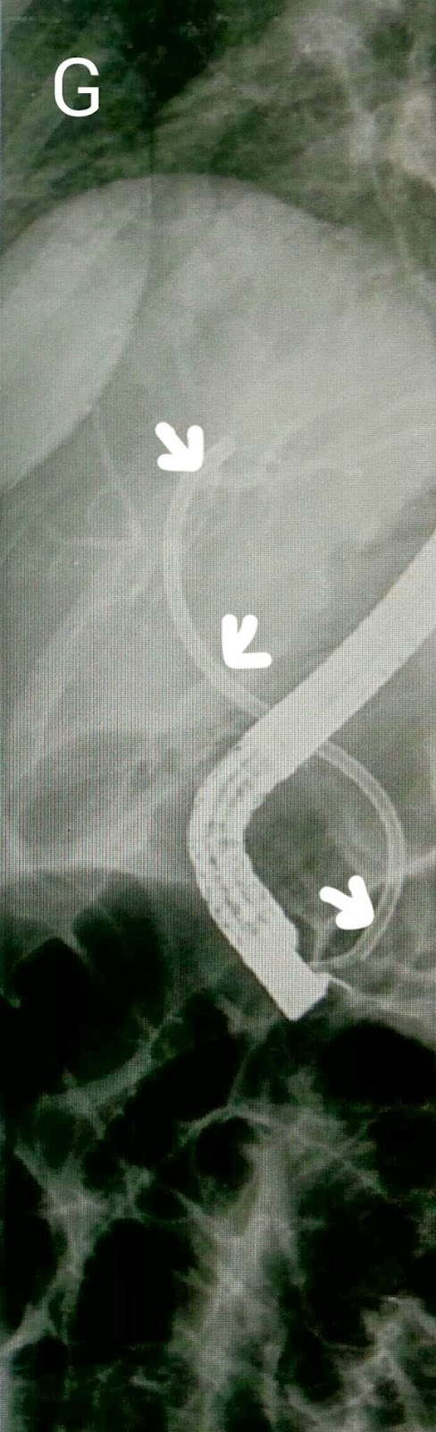
This 18 year girl had developed pancreatitis, most likely biliary in origin and followed by the development of ascites. She was in a a lot of pain and her MRCP showed a significantly dilated pancreatic duct (A: red arrows) with a stone or sludge collection near the terminal end (A: white arrow). The duct was also quite tortuous about a third of the way up (A: blue arrow). Her CT findings also showed the same with a dilated CBD of over 10mm diameter in its terminal portion (B1: black arrow) with an impacted stone/debris collection in its distal end (B2: black arrow). This ERCP was done under propofol sedation due to significant pain reported by the patient. CBD cannulation was quick and contrast injection confirmed the MRCP report with impacted terminal end debris (C: white arrow), dilated pancreatic duct (C: red arrows) and tortuosity (C: blue arrow). We did a pancreatic papillotomy (D1) and the constituency of the impacted material was evident as a white gooey material (D2: white arrow shows debris on the cutting element of papillotome). Further contrast was injected to online the tail region of the pancreatic duct and to rule out any leakage (E)-none was seen.   The papillotomy was extended and further debris was extruded by the extended papillotome (F: white arrow). The pancreatic duct was then swept with balloon (G: white arrow shows the inflated balloon in the pancreatic duct). A large amount of this white gooey debris was removed with repeated balloon sweeps (H1 & H2). Final contrast injection showed a clear pancreatic duct. We decided not to place a stent keeping in mind the large papillotomy done. 

Thursday, January 2, 2014

Bile duct injury and calculi post cholecystectomy

This 35 year old lady had undergone a laparosocpic cholecystectomy at another hospital three months ago. She had been re-admitted with an acute abdomen and the surgeons reported a rent in the CBD and placed a T-tube. She was then referred to us for CBD stenting. Her T-tube cholangiogram showed leakage of contrast (A: white arrow) and what appeared to be a stone (A: red arrow). Contrast injection on ERCP confirmed the cholangiogram findings and showed both the leak and the stone (B: white and red arrows respectively). Curiously, the T-tube was not outlined and we Suspected that it may have gotten dislodged. We did a wide papillotomy (C) and swept the CBD with a stone extraction balloon (D) which yielded a stone (E: white arrow). An occlusion cholangiogram an inflated balloon (F: white arrow) showed a clear CBD. We ended the procedure by placing a 12 cm long single pigtail stent of 10 French diameter (G: white arrows). Single pigtail stent being necessary to anchor the stent inside the CBD keeping in mind the wide papillotomy that had been done. 

Friday, December 27, 2013

One for the Stonehenge: An unusually large CBD stone

This 35 year old lady was one of our staple cases: CBD clearance before undergoing cholecystectomy. The ultrasound report mentioned multiple CBD stones. Contrast injection on ERCP seemed to confirm the ultrasound report with a long line of negative shadows in the CBD (A: white arrow). Once we had done a papillotomy and proceeded to retrieve some of the stones with extraction balloon, we realized that we were dealing with one very large stone. It just kept coming and coming (B & C: white arrows. Blue arrow indicated the balloon). All that was left was a very wide open papilla (D: white arrow) and further balloon sweeps were clean. The calculus filled the duodenal lumen (E: white arrows). Reminds me of the menhirs from the quarry of a certain indomitable Gaul.

Sunday, September 15, 2013

Post cholecystectomy bile duct calculi in a 41 year old gentleman

This patient had undergone cholecystectomy three months earlier and was admitted with obstructive jaundice. Ultrasound showed a 4mm calculus into he distal CBD. ERCP outlined what appeared to be stones into he CBD (A:green arrow). After papillotomy, the CBD was swept with extraction balloon. Occlusion cholangiogram (with inflated balloon in the distal CBD and injection of contrast) showed a clear CBD (B).

Thursday, September 12, 2013

Bile duct stones

We went through a fleshy ampulla (A). The cholangiogram showed a significantly dilated CBD (B) . After a papillotomy, five stones of this type (C) were retrieved via extraction balloon.

Second (actually 3rd) ERCP for post transplant biliary leak

This 60 year old gentleman had earlier undergone ERCP and stenting for an anastomotic biliary leakage a few months earlier http://ercp365.bl...