I am a gastroenterologist. This is a blog of the ERCPs and related endoscopic procedures carried out at my department. Dr Adnan Salim.
Friday, April 25, 2014
Extra large diverticulae flanking ampulla
Lost to follow up
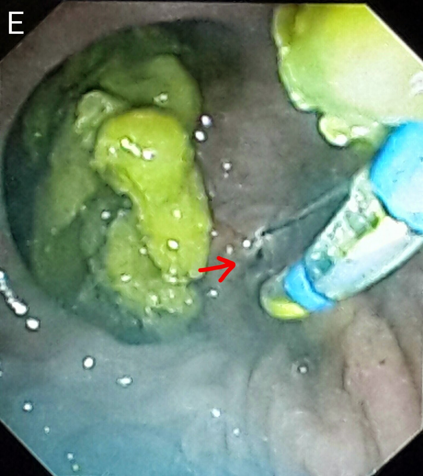
This 45 year old gentleman had suffered from bile duct injury whilst undergoing an open cholecystectomy 10 months ago. The surgeons placed a drain (A: red arrow shows the drain. White arrow shows the large leak-almost looking like the gallbladder in shape. Blue arrow shows the main bile duct). An MRI was done soon after and confirmed the leakage (B: red arrows show the bile leak). He was reffered to our facility and we had placed a 10 French 10cm long plastic stents to bridge the leak. (C: white arrows show the stent) . This was about ten months ago. He was then lost to follow up. He resurfaced at our outpatient out of curiosity asking what was to be done about the Stent we had placed. When questioned as to why he hadn't turned up a month after the procedure, he said he had been told by his physician that his condition has been cured and he needs no more treatment. The moment we went in for ERCP, we were greeted by a normal ampulla with no sign of his placed stent. We thought it had migrated inwards but that wasn't the case as it didn't show up on flouroscopy either. One thing we noted was almost absent bike flow from the ampulla and normal flow from another opening just above it (D: Blue arrow marks the ampulla and white arrow indicates the second opening above it with smeared bile). This second opening was obviously a fistula. Cannulation and contrast injection showed no bile leakage (E: white arrows show the bile duct. Even the fistulous track wasn't outlined). We concluded that the Stent had slipped out and his bole was driving through the fistula. The procedure was ended by a sphincterotomy which was extended to include the fistulous opening above the ampulla (F: white arrow).
Thursday, March 6, 2014
Uncommon biliary tree anatomy
This 54 year old lady had been referred for management of choledocholithiasis prior to undergoing a cholecystectomy. The ultrasound report suggested a single stone in the mid CBD. Initial contrast injection outlined the stone (A: white arrow). Things were about to get interesting, however as further contrast made the picture clearer. We could now see the stone (B: white arrow), the cystic duct (B: blue arrow) and what appeared to be a dividing line right in the middle of the CBD (B: red arrow). And even more contrast finally showed us what we were dealing with: double common bile duct with a common opening with the stone in the left stem (C: white arrow), right and left hepatic ducts draining separately into right and left bile ducts (C: yellow arrows), the cystic duct draining into the right bile duct (C: blue arrow). The arrowheads show black areas which are actually bile ducts viewed head on (C: red arrowhead shows the entrance of the cystic duct into the right bile duct). The stone was removed with an extraction balloon after sphincterotomy (D). The last diagram (E) shows the the modified classification of extrahepatic bile duct duplication as proposed by Choi et al.. Ours seems to be a Va type. The important thing here is the clear communication of this abnormal ductal anatomy to the surgeon who will be doing her cholecystectomy so as to prevent any inadvertent ductal injury and compromised drainage of any of the hepatic lobes.
Friday, February 7, 2014
A balloon for every stone
This 50 year old lady had undergone cholecystectomy twenty years earlier. She had now been having recurrent bouts of right hypochondrial pain. An ultrasound scan showed a 12mm stone impacted in the distal CBD. We were greeted by a big fat papilla with the orifice at its lower end (A: white arrow). Multiple attempts to cannulate the CBD were unsuccessful. We decided to do a precut with a needle knife (B). It was a textbook precut and the CBD opening was nicely exposed (C: white arrow shows the CBD opening - somehow appearing suspiciously similar to the Superman insignia). Contrast injection showed two stones, one below the scope and another just above it (D: white arrows). The papillotomy was extended (E) , however the size was insufficient considering the size of the stones. Our next step was to do a sphincteroplasty using an 18mm diameter TTS balloon (F: white arrow shows the waist of the inflated balloon at the ampulla). Following this, two stones were successfully extracted (G1 & G2). A final cholangiogram with an inflated balloon just above the ampulla showed a duct clear of stones (H).
Wednesday, October 30, 2013
Post cholecystectomy remnant CBD stones
This 38 year old lady had developed obstructive jaundice a few months after undergoing cholecystectomy. The ultrasound report indicated a single stone in the distal CBD. Her cholangiogram outlined a single large stone seen as a negative shadow just above the scope (A: white arrow). The surgical clips from her cholecystectomy were also visible (A: green arrow). We did a papillotomy and we're doing a routine check of adequacy of the papillotomy by withdrawing a fully open sphincterotome. That apparently was enough to pull the stone out (B: white arrow shows the stone and the sphincterotome in the duodenal lumen). We did a routine sweep of the CBD with an extraction balloon, followed by an occlusion cholangiogram with contrast injection over a fully inflated balloon in the distal CBD (C: white arrow shows the inflated balloon) which showed no negative shadows.
Tuesday, September 24, 2013
Cirrhosis of liver with biliary stricture
The patient was a 50 year old lady who had a history of pruritus and liver disease. Viral serology, metabolic profile and autoimmune profiles were normal. Lately she developed recurring jaundice with raised ALP. MRCP showed a distal CBD stricture. On ERCP, intrahepatic channels were poorly outlined on dye injection-the "pruned biliary tree"(red arrow). The CBD showed distal tapering (red arrow). We did a sphincterotomy. That was sufficient to drain the biliary system. The negative shadows in the CBD were only air bubbles (green arrow).
Second (actually 3rd) ERCP for post transplant biliary leak
This 60 year old gentleman had earlier undergone ERCP and stenting for an anastomotic biliary leakage a few months earlier http://ercp365.bl...
-
This 22 year old lady had undergone living donor liver transplant at our centre for hepatitis B related liver disease. She developed an ana...
-
This gentleman was referred from another hospital. Cholangiogram showed a distal CBD stricture (A:green arrow). We placed a 10Fx10cm plastic...
-
A 50 year old lady had developed obstructive jaundice over the past few months and had been admitted with recent onset of fever. She was fou...