Showing posts with label MRCP. Show all posts

Saturday, September 12, 2015

Second follow up ERCP for post liver transplant anastomotic biliary stricture (Case K)

This 22 year old lady had undergone living donor liver transplant at our centre for hepatitis B related liver disease. She developed an anastomotic stricture shortly after her surgery and underwent an ERCP in April of this year (A 7 French stent of 15 cm length was initially placed after dilatation of stricture with graduated dilatation catheters but she developed GI bleeding the same day. On endoscopy done the next day, the distal end of the stent was seen to erode the opposite duodenal wall and cause ulceration. This was removed and a shorter stent of the same diameter but shorter 12 cm length was placed). Now, she had developed pruritus and fever. Her stent was removed (A). The next image shows her MRCP which was done prior to her first ERCP (B: White arrow shows the anastomotic stricture site and red arrow shows a collection next to the biliary tree). Contrast injection after stent removal showed the narrowed stricture area (C: Black arrow) and what appears to be a sludge ball above the stricture (C: White arrow). A stricture dilatation balloon of 40 mm length and 6 mm diameter was used to dilate the stricture ( D: Black arrow). A stent assembly was placed across the affected area (E: Black arrow) and a 7 French plastic stent of 12 cm length was deployed (F: Black arrows).


Wednesday, April 8, 2015

Post liver transplant anastomotic biliary stricture (CASE H)

This 42 year old gentleman had undergone a non related living donor liver transplant at a foreign centre one year ago (March 2014). For the past 6 weeks he had been complaining of pruritus and clay coloured stools. Liver biochemistry revealed rising ALP and bilirubin levels. His MRCP showed a subtle anastomotic stricture (A: red arrow) and prominent intrahepatic biliary channels (A: blue arrows). Contrast injection on ERCP showed the stricture (B: red arrow0 but the guidewire kept going into a sharp bend (B: white arrow) and kept curling up on itself (C: white arrow). We were finally able to get our guidewire across (D: black arrows) whilst avoiding the bend. Our papillotome was then pushed up along a mightily curved path (E: black arrow). The track was first dilated using a 7 French graduated dilatation catheter (F1: black arrows show the catheter while the red arrow indicates the level of stricture). This was followed by a larger 8.5-10 French catheter (F2 & F3: black arrows. Note: Star Wars fans will notice the uncanny resemblance of this catheter's "mouth" to Greedo's). A 7 French stent of 15 cm length was planned to be deployed. This went south as we weren't able to push the stent beyond even the first curve of the hairpin (G: white arrows show the failed stent deployment proximal to the stricture). No fear! We went again with aching muscles and did a repeat dilatation of the track with our  10 French "Greedo" catheter (H: black arrows show the catheter snaking up and red arrow marks the stricture site). A 7 French stent of 12 cm length was deployed. 
Note: This exercise highlights the difficulty while traversing a stricture using  smaller 7 French stent as these are deployed directly over the guidewire without the assembly. No assembly means less stiffness and strength which in turns translates to the stent bending before rather than pushing through an area of resistance.


Tuesday, February 3, 2015

To boldly drain what others have not drained before

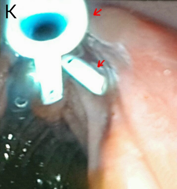
This 26 year old lady had a history of recurrent cholangitis. She had extra hepatic portal venous obstruction with varies at her porta causing compression of the extra hepatic biliary channels. She had previously undergone three ERCPs. The second ERCP showed her dilated left and right biliary systems (A: black arrows) with luminal narrowing of the confluence, CHD & CBD (A: white arrows). Following dilatation with a graduated dilatation catheter, a 10 French plastic stent had been placed in her right system (B: black arrows) but her left system could not be cannulated (B: white arrows). On her 3rd ERCP, the stent had been removed. Now, 4 months later, she presented with cholangitis. Her MRCP images showed the same findings along with stones on the CBD (C: red arrow marks the calculi, black arrows show the narrow CBD, white arrow indicates the narrowing at the hilum and blue arrows show the dilated right and left ductal systems). We booked her for her 4th ERCP. Our guidewire was first manoeuvred into her left system (D: black arrow shows the guidewire and white arrow shows the dilated left system). This was followed by dilatation of the track using a 10 French graduated dilatation catheter (E: white arrows show the dilatation catheter). We then swept the CBD with a biliary balloon (F:black arrows mark the balloon assembly in the CBD) and a  moderate amount of small calculi and sludge was removed (G1: red arrows mark the removed calculi. G2: black arrow shows the "clean sweep" ampulla). We then passed another guidewire in the right system (H: black and white arrows show the two guide wires in the right and left systems, respectively). A 10 French plastic stent of 12 cm length was passed into the right system (I: black arrows mark the stent in the right main duct). A thinner 7 French stent (also of 12 cm length) was then passed into the left system (J: white arrows show the stent in the left main duct while black arrows show the first stent in the right system. K: red arrows show the two stents draining happily in the duodenal lumen).

Friday, May 9, 2014

Major biliary tree disruption post surgery

This 52 year old lady had been referred from another hospital following bile duct injury during open cholecystectomy. She underwent a repeat laparotomy after her gall bladder surgery for the leak. A drain was placed in the sub hepatic space and the patient was subsequently sent to us. Her MRCP wasn't the best of images we had seen, with major motion artifacts (problem with holding her breath, we suppose). A disruption in the bile duct (A: red arrow) and leakage (A: blue arrow) was clear. Contrast injection after CBD cannulation showed the extent of damage, with structures, leaks and what appeared to be an uncommon  anatomical variant of the biliary tree (B: green arrows show the leak. Blue arrow shows a major stricture. Black & white arrows show what appear to be right and left hepatic ducts but without the classical confluence morphology we're used to seeing. Red arrow indicates a 0.035 guidewire which has been placed into the right system). The right system,where we had managed to place the wire, was dilated with a 10 French graduated dilatation catheter (C: black arrows). We then left one wire in the right duct and made multiple attempts to place the second wire into the left duct in order to dilate it (D1 to D4: black arrow shows the first wire in the right system and white arrow shows the second wire as we attempt to place it in the left duct). Alas we were unsuccessful. A decision was made to stent the right duct. A stent assembly was then passed. As we passed the stent over it, the force required to pass the strictured segment (despite dilatation done earlier) caused the stent assembly to warp into the area of leakage (E: white arrows show the stent assembly and black arrow shows the assembly bent into the leak area). A 10 French stent of 12 cm length was then deployed (F1 &  F2: white arrows). 


Friday, April 25, 2014

Lost to follow up

This 45 year old gentleman had suffered from bile duct injury whilst undergoing an open cholecystectomy 10 months ago. The surgeons placed a drain (A: red arrow shows the drain. White arrow shows the large leak-almost looking like the gallbladder in shape. Blue arrow shows the main bile duct). An MRI was done soon after and confirmed the leakage (B: red arrows show the bile leak). He was reffered to our facility and we had placed a 10 French 10cm long plastic stents to bridge the leak. (C: white arrows show the stent) . This was about ten months ago. He was then lost to follow up. He resurfaced at our outpatient out of curiosity asking what was to be done about the Stent we had placed. When questioned as to why he hadn't turned up a month after the procedure, he said he had been told by his physician that his condition has been cured and he needs no more treatment. The moment we went in for ERCP, we were greeted by a normal ampulla with no sign of his placed stent. We thought it had migrated inwards but that wasn't the case as it didn't show up on flouroscopy either. One thing we noted was almost absent bike flow from the ampulla and normal flow from another opening just above it (D: Blue arrow marks the ampulla and white arrow indicates the second opening above it with smeared bile). This second opening was obviously a fistula. Cannulation and contrast injection showed no bile leakage (E: white arrows show the bile duct. Even the fistulous track wasn't outlined). We concluded that the Stent had slipped out and his bole was driving through the fistula. The procedure was ended by a sphincterotomy which was extended to include the fistulous opening above the ampulla (F: white arrow). 


Tuesday, April 1, 2014

Long CBD stricture secondary to extrinsic compression

This 54 year old lady had a dilated gallbladder compressing the mid CBD upto the confluence (A: MRCP & B: CT scan ; Red arrows showing the dilated gallbladder. Blue arrows show the compression at the confluence and green arrows show dilated intrahepatic channels. C: CT scan; Red arrows marking the gallbladder). Contrast injection on ERCP outlined the narrowing in the CBD (D: black arrows) and dilated intrahepatic ducts. A stent assembly was placed across the affected segment (D). We passed a 15 cm plastic stent of 10 French diameter (E: white arrows show the stent loaded on its assembly. F: Black arrows show the stent passed across the structured segment and into the dilated intrahepatic channels)

Second (actually 3rd) ERCP for post transplant biliary leak

This 60 year old gentleman had earlier undergone ERCP and stenting for an anastomotic biliary leakage a few months earlier http://ercp365.bl...