Showing posts with label cholelithiasis. Show all posts

Friday, April 25, 2014

Extra large diverticulae flanking ampulla

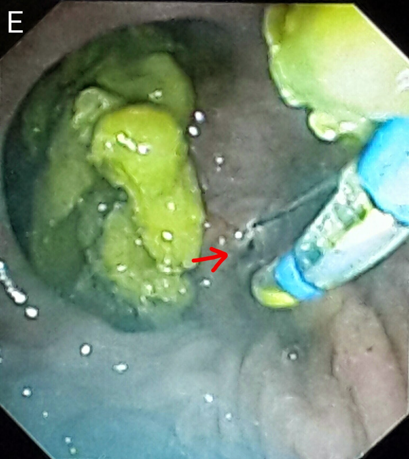
This 60 year old gentleman had been referred following the development of classical obstructive jaundice signs. His ultrasound reported an ampullary mass lesion. The CT report showed a greatly distended CBD with a large calculus at the terminal end (A: white arrow posts to the stone and black arrows delineate the CBD). There was a "whoa" moment the moment the papilla came into view as it was flanked by two huge diverticulae stuffed with food (B: red arrows. White arrow points to the papilla). The scope kept slipping and cannulating the ampulla was trickier than expected (C). Contrast injection showed a dilated CBD with a large stone near the lower end (D: black arrow). We did a small sphincterotomy (E: red arrow). A 10 French plastic stent of 10 cm length was then deployed (F1: red arrow shows the stent and blue arrow points to the stent assembly. F2: black arrows show the stent). The patient was referred to the surgeon for cholecystectomy and bile duct clearance. 

Thursday, March 6, 2014

Uncommon biliary tree anatomy

This 54 year old lady had been referred for management of choledocholithiasis prior to undergoing a cholecystectomy. The ultrasound report suggested a single stone in the mid CBD. Initial contrast injection outlined the stone (A: white arrow). Things were about to get interesting, however as further contrast made the picture clearer. We could now see the stone (B: white arrow), the cystic duct (B: blue arrow) and what appeared to be a dividing line right in the middle of the CBD (B: red arrow). And even more contrast finally showed us what we were dealing with: double common bile duct with a common opening with the stone in the left stem (C: white arrow), right and left hepatic ducts draining separately into right and left bile ducts (C: yellow arrows), the cystic duct draining into the right bile duct (C: blue arrow). The arrowheads show black areas which are actually bile ducts viewed head on (C: red arrowhead shows the entrance of the cystic duct into the right bile duct). The stone was removed with an extraction balloon after sphincterotomy (D). The last diagram (E) shows the the modified classification of extrahepatic bile duct duplication as proposed by Choi et al.. Ours seems to be a Va type. The important thing here is the clear communication of this abnormal ductal anatomy to the surgeon who will be doing her cholecystectomy so as to prevent any inadvertent ductal injury and compromised drainage of any of the hepatic lobes.

Friday, December 27, 2013

One for the Stonehenge: An unusually large CBD stone

This 35 year old lady was one of our staple cases: CBD clearance before undergoing cholecystectomy. The ultrasound report mentioned multiple CBD stones. Contrast injection on ERCP seemed to confirm the ultrasound report with a long line of negative shadows in the CBD (A: white arrow). Once we had done a papillotomy and proceeded to retrieve some of the stones with extraction balloon, we realized that we were dealing with one very large stone. It just kept coming and coming (B & C: white arrows. Blue arrow indicated the balloon). All that was left was a very wide open papilla (D: white arrow) and further balloon sweeps were clean. The calculus filled the duodenal lumen (E: white arrows). Reminds me of the menhirs from the quarry of a certain indomitable Gaul.

Monday, October 21, 2013

CBD stone removal pre cholecystectomy

A 50 year old lady had come for CBD stone removal before undergoing cholecystectomy. The MRCP showed a stone in the distal CBD (A: green arrow)  and gallbladder calculi (A: red arrow) and a dilated CBD (A: yellow arrow). Contrast injection on ERCP showed the distal CBD stone as in the MRCP and another negative shadow above the ERCP scope (B: green arrows marking the stones and yellow arrow showing a dilated CBD). The distal stone was removed by the papillotome (C: green arrow shows the stone and red arrow marks the papillotome). The CBD was swept with balloon (D: green arrow shows the inflated balloon) and the second stone was also removed (E: green arrows show both the stones in the duodenal lumen).

Monday, October 7, 2013

CBD stone removal prior to cholecystectomy

A 35 year old lady with cholelithiasis and choledocholithiasis, Sent by the surgical team for CBD clearance prior to cholecystectomy.
On ERCP, contrast injection showed a single stone in the CBD as a rounded negative shadow (A : green arrow). We did a wide papillotomy (B: green arrow) followed by removal of the stone with a biliary balloon (C: green arrow points to the inflated balloon & D: green arrow shows the removed stone in the duodenal lumen).

Saturday, October 5, 2013

Bile duct calculi in a patient with multiple co-morbids

This 66 year old gentleman took the cake for multiple coexisting diseases in addition to his biliary pathology. He had diabetes, hypertension, ischemic heart disease, epilepsy and depression. He also had cholelithiasis and choledocholithiasis. We engaged the papilla (A:green arrow) but couldn't cannulate the CBD. We resorted to a precut with a needle knife (B:green arrow) which clearly identified the CBD opening (C: green arrow). Contrast injection showed stones in the distal CBD (D: green arrow). Our patient became distressed and felt pain during extension of papillotomy (which was quite unusual) by this time and we quickly ended the procedure by cancelling stone extraction and placing a plastic stent of 10 French diameter and 10cm length (E: green arrows). In our opinion, the papilla seemed unhealthy. This, plus the transaminase and ALP levels in the high hundreds prompted us to advise a CA 19-9 and abdominal CT scan before a repeat ERCP for stone extraction (to be done under propofol sedation)

Thursday, October 3, 2013

Pancreatitis with pancreatic and bile duct calculi

This 48 year old gentleman had a history of recurrent pancreatitis. MRCP showed calculi in the distal and middle pancreatic duct (A: green arrows) and calcification in the region of pancreatic head (A: red arrows). The ultrasound report indicated the presence of small calculi in the distal CBD in addition to the pancreatic duct calculi. On ERCP, the CBD was cannulated, followed by contrast injection which did not show any stones. The CBD showed distal tapering (B: green arrows - which may be due to pressure from the calcified pancreatic head). We did a papillotomy followed by sweep with balloon which also didn't result in any stone or sludge. Despite repeated attempts, we couldn't cannulate the pancreatic duct. The case has now been rescheduled to a later date. 

Wednesday, September 25, 2013

Bile duct clearance with an intra-diverticular ampulla

Have a close look at the first image (A1: green arrow). The ampulla is inside a seemingly small diverticulum. However, these diverticuli change their shape considerably with bowel movements. Since this was a pre-cholecystectomy CBD clearance, when we saw the ampulla, we were skeptical about attempting stone extraction (the patient's bilirubin was normal) and thought to advise the surgeon to clear the CBD as well. In the second image (A2: green arrow), the ampulla seems to be pouting out. The diverticulum was invaginating and evaginating with bowel movements. Contrast injection showed a single large stone (B: green arrow). We did a papillotomy (A2) and removed the stone with extraction balloon. Repeat contrast injection showed a clear CBD (C & D).

Wednesday, September 18, 2013

Choledocholithiasis and cholelithiasis

A middle aged lady with gallbladder and bile duct stones. The ultrasound mentioned a single 12 mm calculus. An interesting thing was the presence of a biliary fistula superior to the papilla (A:green arrow). Contrast injection showed multiple calculi (B). We did a wide papillotomy (C). The stones were removed using an extraction balloon (D1 & D2). Post stone removal cholangiogram showed a clear CBD (E).

Tuesday, September 17, 2013

Procedure related complication: Pre cholecystectomy CBD clearance

This patient was an obese 35 year old lady with systemic hypertension and heart disease. She had been referred from another facility for ERCP and removal of CBD stones prior to undergoing a cholecystectomy. The case turned out to be a tough one. We were able to cannulate the pancreatic duct and went for a transpancreatic papillotomy. CBD cannulation followed. Surprisingly, there weren't any stones inside (A: green arrow & B) neither did the ampulla look like a stone had passed through it.
The CBD showed a short stricture in its terminal part. We passed a plastic stent (C: green arrow)and admitted her for overnight monitoring as she had gotten quite restless (the procedure wasn't being done under propofol sedation) and her blood pressures had shot up. The patient was underwent an abdominal x ray which was normal. A CT scan however showed free air in the retroperitoneal space (D: red arrows). She was afebrile and bowel movements were normal. It was decided to manage her conservatively. She was improving 96 hours later. 

Second (actually 3rd) ERCP for post transplant biliary leak

This 60 year old gentleman had earlier undergone ERCP and stenting for an anastomotic biliary leakage a few months earlier http://ercp365.bl...