Wednesday, August 28, 2013

Biliary stent penetrating opposite duodenal wall

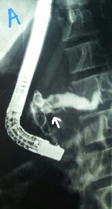
Not a common sight. We had gone for stent removal and attempt stone extraction (the patient had previously had cholangitis and stone extraction was incomplete. A plastic stent had been placed). The stent had penetrated the opposite duodenal wall, a closed perforation. We had to wrestle a bit with the forceps, pushing the stent back into the CBD. It was then removed with a snare.

Monday, August 26, 2013

Intra-diverticular papilla

Thought it would be a simple "Snatch and Grab"  bile duct stone. Turned out this patient had an intra-diverticular papilla (like one of our previous transplant patients). Cannulation took its sweet time. One truly appreciates a normal papilla in such cases.

Sunday, August 25, 2013

Gastric outlet obstruction

This 52 year old lady was referred for achalasia balloon dilatation from another hospital where an upper GI endoscopy had also been done and was reported to be unremarkable. Her esophageal manometry suggested achalasia of the cardia. We went in with a plan for balloon dilatation of the lower esphageal sphincter. However, extensive infiltration was seen at the cardia (A) and in antral area (B). Dilatation was, of course,  cancelled. We took biopsies and advised a biphasic CT of the chest and abdomen. The important lesson was that even when going for an achalasia dilatation in a fully worked up case, always have a good look at the cardia to rule out pseudoachalasia. 


Pancreatic duct calculi

This 12 year old girl had a history of multiple episodes of pancreatitis secondary to pancreatic ductal calculi. On ERCP the pancreatic duct was cannulated followed by contrast injection which initially outlined negative shadows in the head region (A: white arrow). However further contrast injection showed the duct full of calculi (B: white arrows). We didn't attempt to remove. The patient was referred to the surgical team for further management. 

Esophagopleural fistula

This patient had an esophagopleural fistula secondary to a gunshot wound. The bullet track was from the right shoulder (A: red arrow shows the entry point and dotted lines show the track), through the mediastinum (miraculously missing any vital organs) and finally embedded in the left shoulder (A: green arrow). This resulted in a right pleural effusion for which a drain was placed (B: White arrow shows the drain. Green arrow shows the bullet embedded in the left shoulder). Endoscopy showed the fistula clearly for which a 23mm x 155mm self expanding covered metallic stent was placed (C: Red arrow shows the stent assembly and green arrow shows the fistula opening. D: white arrows show the deployed stent). The patient was reviewed after three months and some polypoid growths were seen near the upper and lower uncovered areas of the stent (E). The stent was removed with forceps (F). A post removal gastrograffin swallow showed no leakage of contrast material (G1 to G4: green arrows in G1 and G4 show the bullet still lodged near the left shoulder). 


Second (actually 3rd) ERCP for post transplant biliary leak

This 60 year old gentleman had earlier undergone ERCP and stenting for an anastomotic biliary leakage a few months earlier http://ercp365.bl...