Friday, September 26, 2014

Third follow up ERCP for post transplant anastomotic stricture (CASE A)

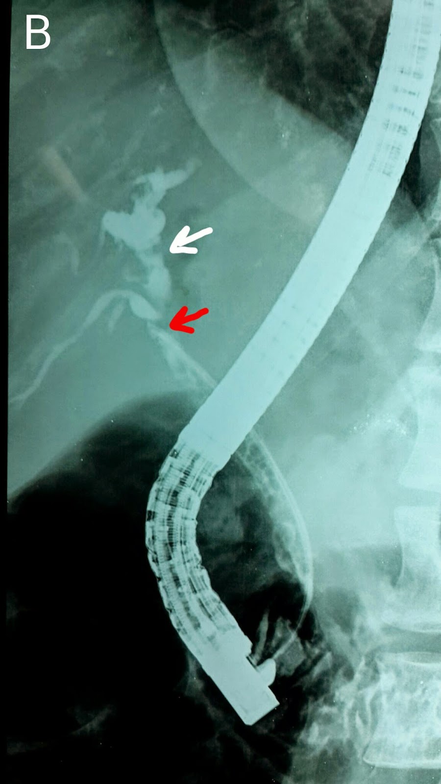
We last saw this 50 year old gentleman two and a half months earlier when we dilated his anastomotic stricture with a 6mm balloon and placed a 10 French stent http://ercp365.blogspot.com/2014/09/second-follow-up-ercp-for-post-liver.html. His bilirubin levels had started to rise and we expected a blocked stent. His ultrasound confirmed this with no pneumobilia. We removed his previous stent using a snare (A: White arrows mark the wires of the snare around the stent. Blue arrow marks the stent. A guidewire was manoeuvred across the stricture (B: Black arrow shows the level of the stricture on the guidewire). Contrast injection again confirmed the narrowing (C: Black arrows show the narrowing at the anastomotic site). We decided to go for a bigger balloon this time and an 8mm diameter balloon was placed across the stricture (D: White arrows). The balloon was inflated (E: White arrows show the inflated balloon with a "waist" formation at the specific point of concern). Following dilatation, repeat contrast injection showed adequate dilatation (F: Black arrows now show no narrowing).  A stent assembly was then placed (G: White arrows). A 10 French plastic stent of 15 cm length was deployed.

Second follow up ERCP for post liver transplant anastomotic stricture (CASE A)

This was a 50 year old gentleman who had undergone living donor liver transplant in June 2013. He underwent his first ERCP after developing an anastomotic stricture in February. An 8.5 French stent of 12cm length was placed after dilating the stricture with graduated dilatation catheters http://ercp365.blogspot.com/2014/02/post-transplant-anastomotic-biliary.html. We removed his previously placed stent. Contrast injection outlined a persistent stricture (A: Black arrow). We dilated the stricture using a 6 mm balloon of 3 cm length (B: White arrows). A 10 French plastic biliary stent of 15 cm length was placed (C1 & C2: White arrows).

Saturday, September 20, 2014

Fifth follow-up ERCP for post transplant biliary stricture (CASE B)

This was this 24 year old's fifth follow up visit. We removed the stent placed in his last visit http://ercp365.blogspot.com/2014/09/fourth-follow-up-ercp-for-post.html. Contrast injection showed a persistent stricture (A: Black arrows). We used a 10mm stricture dilatation balloon of 3cm length to dilate the stricture (B & C: Black arrows show the inflated balloon and red arrow marks the "waist"of the balloon at the strictured segment in B). Marked improvement in the previously narrowed site of ductal anastomosis was noted (D: Black arrows).

Fourth follow-up ERCP for post transplant biliary stricture (CASE B)

This 24 year old gentleman was undergoing his fourth ERCP this patient was undergoing for his anastomotic stricture. No stent was placed at his last visit http://ercp365.blogspot.com/2014/03/third-follow-up-ercp-for-post.html but this time around he was developing fever and slight rise in his bilirubin levels. His ampulla showed adequate outflow of bile (A: Black arrow). Initially our guidewire passed only up to just below the anastomotic site and contrast outlined a normal calibre lower CBD (B: Black arrows indicate the native bile duct). Finally our guidewire crossed the stricture site and the graft ducts were also outlined (C: White arrows show the stricture. Blue arrow shows the guidewire which isn't yet going straight up as we would like. Red arrow shows the dilated ducts in the graft). With repeated efforts we were able to get the wire go up in a direct line (D: Black arrows show the native bile duct, of normal calibre. White arrows show the structured anastomotic site. Blue arrow shows the tip of the guidewire). We dilated the stricture with a 10 French graduated dilatation catheter (E: White arrows mark the catheter). A stent assembly was then placed across the stricture and a 10 French plastic stent of 12 cm length ( F: White arrows show the stent assembly and black arrows indicate the stent passing over the assembly) was passed (G: Black arrows mark the deployed stent).

No one gets left behind-Retained bile duct stone post cholecystectomy

This 32 year old lady had undergone open cholecystectomy at a peripheral centre. Unfortunately, CBD clearance was incomplete and she was referred to us with a large calculus in the left main duct. She was accompanied by a T-tube cholangiogram (A: Red arrow shows the stone and white arrows shows the T-tube). Before going in, we did a repeat cholangiogram via the T-tube and it showed the stone had migrated to the distal CBD (B: Red arrow shows the meniscus sign, indicating the stone). The T-tube was removed and contrast injection following wire cannulation of the CBD showed that the stone had become lodged in a "recess" of sorts (C: Red arrow), made when the CBD was kinked by the pull of the T-tube. Seeing the considerable size of the stone, we decided to do as wide  a papillotomy as possible (D: White arrow). First weapon of choice was our trusty biliary balloon (E: Black arrow shows the inflated balloon proximal to the stone. Red arrow shows the stone itself. White arrow indicates the marked kink that had developed in the CBD once the T-tube was pulled out). The balloon kept slipping by the side of the stone. Next up was the dormia basket (F: White arrow). The basket was manoeuvred past the stone (G1: Black arrows show the open basket and red arrow marks the stone). The "perp" was finally "apprehended (G2: White arrows show the basket and red arrow shows the captured stone). The stone was pulled out (H: Red arrow shows the stone and black arrows indicate the basket wire around it. I: The retrieved stone seen on a gauze-Homo Sapiens forelimb thumb for size comparison. Oversized glove is evident).

Second time lucky-Managing metastatic peri-ampullary tumor with cholangitis


This 65 year old lady had been admitted with obstructive jaundice and cholangitis secondary to a metastatic peri-ampullary tumor. We encountered a swollen ampulla on duodenal intubation (A). Cannulation and contrast injection outlined a significantly dilated pancreatic duct (B: Black arrows) and what appeared to be the accessory duct (B: White arrow). We did a pancreatic papillotomy (C: White arrow). Further attempts here were unsuccessful and slight scope with-drawl showed an opening at the upper surface of the ampulla seen in image A (D: Black arrow), which was cannulated (E: Black arrow). This was the proper route as evidenced by wire cannulation (F: Black arrows). Contrast injection in this area outlined a dilated proximal bile duct and intrahepatic channels (G: White arrows) with no distal or mid duct delineation. A 10 mm diameter and 8 cm long partially uncovered metallic stent was prepped (I. We used a Micro-Tech stent. These come with an internal stiffening wire with a ring for removal prior to insertion: Red arrow). The stent assembly was placed across the strictured segment unto the dilated duct (J: Black arrows). The stent was then deployed (K1: The stent being deployed. Red arrow marks the gold reference mark which is kept visible just at the edge of field of view. K2: The deployed stent. A gush of contrast and white pus was seen. L1 & L2: White arrows show the fully deployed stent with black arrows indicating the proximal and distal radiographic markers).


Fifth follow up ERCP for post cholecystectomy biliary stricture.

This was this 40 year old lady's fifth ERCP. Somehow her stricture didn't seem to respond to conventional methods. This time around we removed the two stents that were placed in her last visit http://ercp365.blogspot.com/2014/02/fourth-follow-up-ercp-for-post.html (A). Contrast injection showed the strictured area (B: Red arrow) and dilated biliary channels proximal to the stricture (B: White arrow). A 10 French plastic stent of 12 cm length was placed first (C: White arrows), crossing the affected area (C: Red arrowhead). This was followed by a longer 15 cm plastic stent, also of 10 French diameter (D: Blue arrows. White arrows mark the first stent).

Second (actually 3rd) ERCP for post transplant biliary leak

This 60 year old gentleman had earlier undergone ERCP and stenting for an anastomotic biliary leakage a few months earlier http://ercp365.bl...