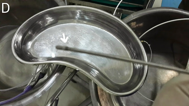
This 60 year old lady had a hilar cholangiocarcinoma. Significant intrahepatic biliary dilatation was evident on contrast injection (A: Red arrows show dilated intrahepatic ducts and black arrows show the affected narrowed hilar area). We placed one guidewire in the right system (B1: red arrows) and attempted to manoeuvre a second one into the left system (B1: black arrows and B2). This was unsuccessful and it kept going into the right system along with the previous wire. The strictured segment was dilated using an 8.5 French graduated dilatation catheter (C1 & C2: black arrows show the catheter and red arrows show the second guidewire). The second guidewire was removed and a stent assembly was pushed into the right system (D: black arrows). A 10 French plastic stent of 10 cm length was placed (E: white arrows). The patent was started on antibiotics and she predictably developed fever. However it was short lived and we were able to discharge her home within the week. Retrospectively, what appear to be left sided channels in "A" may well have been right sided channels as well (appearing so due to the patient's position. These suspected left channels are also free of contrast in "C1", so we might not have injected the left system in the first place.
I am a gastroenterologist. This is a blog of the ERCPs and related endoscopic procedures carried out at my department. Dr Adnan Salim.
Thursday, July 31, 2014
A very large intrahepatic cholangiocarcinoma
This was a 30 year old gentleman who had presented with obstructive jaundice. His CT scan showed a very large mass of approximately 12x11cm occupying most of the left one (A: white arrows) with involvement of the porta hepatis and extensive intra-abdominal lymphadenopathy. On ERCP, contrast injection following placement of guidewire outlined a long stricture involving the region of ductal confluence (B: white arrows) with dilated left sided ducts proximal to stricture (B: red arrows). Since the right system was not outlined, we left our first guidewire in the left system (C: white arrows) and attempted to pass a second guidewire in the right system (C: black arrows). This was unsuccessful. Luckily, no contrast had gone into the right system. We dilated the structured segment using a graduated 9 French dilatation catheter (D1 & D2: white arrows). A 12 cm long plastic stent of 10 French diameter was then deployed (E1 & E2: black arrows )
Stenting a malignant duodenal stricture
This 70 year old gentleman had previously undergone placement of a metallic biliary stent last year for an ampullary tumor. He was subsequently lost to follow up and had only now presented with gastric outlet obstructive symptoms. A swallowed contrast study (A) clearly showed the problem. The previously placed stent was outlined (A: white arrows) along with the intrahepatic biliary channel (A: green arrows). The stricture was also seen as a thin structured segment in the second part of the duodenum (A: red arrow). On endoscopy, the affected area was seen with almost complete luminal obstruction (B: white arrow). We placed a biliary cannulation catheter across the stricture followed by contrast injection which outlined the extent of the stricture (C: red arrowheads) and the unaffected bowel segment beyond the stricture (C: blue arrow). The biliary stent was also seen (C: white arrowheads). A guidewire was then placed across the stricture (D: white arrows) followed by a stent assembly (E: black arrows). A 12 cm long stent of 20 mm diameter was then deployed (F1 & F2 : black arrows show the stent assembly. White arrowheads show the deployed portion of the stent . Red arrowheads show the distal and middle radiographic markers of the stent. F3: Fully deployed stent. Stent outlined with white arrowheads. Red arrowheads showing the radiographic stent markers. Blue arrow showing the previously injected contrast in the lumen of the distal portion of the stent. G1 & G2: The stent as seen by the endoscope: white arrows. Compare with B).
Tuesday, May 20, 2014
Extrinsic biliary compression in a patient with Familial Adenomatous Polyposis
This 35 year old gentleman was a diagnosed case of Familial Adenomatous Polyposis who had developed obstructive jaundice secondary to extrinsic biliary compression. This was evident on MRCP (A: red arrow). This was most likely a lymph node exerting pressure on the bile duct. Contrast injection on ERCP confirmed the MRCP findings (B: red arrow). We dilated the stricture tract with a 10 French dilator (C: black arrows). Further advancement of the dilator revealed a sharp bend (D: white arrow marks the bend. Black arrows delineate the stent). A 12cm long plastic stents of 10 French diameter was placed (E: white arrow shows the "kink" in the stent at the aforementioned bend. Black arrows outline the stent). A snare was used to pull the stent back to straighten it out (F: red arrow shows the snare around the stent. G: black arrows mark the non kinked stent, properly deployed).
Follow up case of post cholecystectomy biliary leak
This 38 year old lady had previously undergone biliary stunting for a bile leak http://ercp365.blogspot.com/2013/12/and-another-bile-leak-following.html which had occurred following cholecystectomy. We had placed a 10 cm long 10 French plastic stent. We removed the stent (A). The sludge coating prompted us to do a biliary sweep with a stone extraction balloon (B: white arrow shows the papilla with a previous sphincterotomy and the balloon assembly being inserted). The CBD was swept (C: black arrows shows the inflated Ballon in the CBD). Gan occlusion cholangiogram with the balloon inflated at the distal end of the CBD was done (D: black arrow shows the inflated balloon. White arrows delineate the CBD with no leakage). The patient was discharged.
Friday, May 9, 2014
Major biliary tree disruption post surgery
This 52 year old lady had been referred from another hospital following bile duct injury during open cholecystectomy. She underwent a repeat laparotomy after her gall bladder surgery for the leak. A drain was placed in the sub hepatic space and the patient was subsequently sent to us. Her MRCP wasn't the best of images we had seen, with major motion artifacts (problem with holding her breath, we suppose). A disruption in the bile duct (A: red arrow) and leakage (A: blue arrow) was clear. Contrast injection after CBD cannulation showed the extent of damage, with structures, leaks and what appeared to be an uncommon anatomical variant of the biliary tree (B: green arrows show the leak. Blue arrow shows a major stricture. Black & white arrows show what appear to be right and left hepatic ducts but without the classical confluence morphology we're used to seeing. Red arrow indicates a 0.035 guidewire which has been placed into the right system). The right system,where we had managed to place the wire, was dilated with a 10 French graduated dilatation catheter (C: black arrows). We then left one wire in the right duct and made multiple attempts to place the second wire into the left duct in order to dilate it (D1 to D4: black arrow shows the first wire in the right system and white arrow shows the second wire as we attempt to place it in the left duct). Alas we were unsuccessful. A decision was made to stent the right duct. A stent assembly was then passed. As we passed the stent over it, the force required to pass the strictured segment (despite dilatation done earlier) caused the stent assembly to warp into the area of leakage (E: white arrows show the stent assembly and black arrow shows the assembly bent into the leak area). A 10 French stent of 12 cm length was then deployed (F1 & F2: white arrows).
Saturday, April 26, 2014
Migrated biliary stent
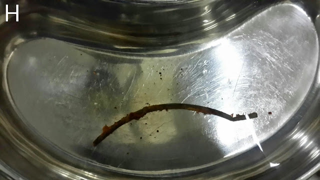
This 40 year old gentleman is an old patient of ours. He has been treated for hepatitis C and extra hepatic portal venous obstruction. He has been regularlyundergoing ERCPs at our center for bile duct obstruction. We usually clear the CBD and place a new plastic stent at each visit. This time around we couldn't see the previously placed stent and suspected that it had migrated into the bile duct. This was confirmed on flouroscopy (A: black arrows). We first attempted to pull the stent out using a stone extraction basket (B: white arrows show the stent. Black arrows mark the basket). This was unsuccessful so our next maneuver was to thread the stent with a guidewire (C: white arrows show the stent and black arrows mark the guidewire). The wire was passed to thread a Soehendra stent retriever over it (D: white arrow). The stent was properly engaged (E: white arrow indicates the stent retriever and black arrow market the engaged lower end of the stent). The old stent was then pulled out (F: black arrow shows the friable sludge ridden end of the stent being pulled by the Soehendra stent retriever-white arrow). We could have directly pulled free stent out through the scope channel but were concerned that it might break since the distal end was very cracked and friable. We used a snare to pull it out (G: white arrow shows the snare. H: The retrieved stent l. A wide papillotomy was done (I: white arrow). The CBD was swept with a stone extraction balloon (J: black arrow shows the inflated balloon in the bile duct). A lot of debris was removed (K: white arrows). Finally a 10 French diameter plastic stent of 12 cm length was placed (L: black arrows). This was a stent with a curved proximal end. Not the the classical pigtail shape. This would help in retention of the stent keeping in mind the papillotomy.
Subscribe to:
Posts (Atom)
Second (actually 3rd) ERCP for post transplant biliary leak
This 60 year old gentleman had earlier undergone ERCP and stenting for an anastomotic biliary leakage a few months earlier http://ercp365.bl...

-
A healthy 48 year old gentleman with a recent history of jaundice. The MRCP outlined a dilated proximal CBD with a small portion of the dist...
-
This 22 year old lady had undergone living donor liver transplant at our centre for hepatitis B related liver disease. She developed an ana...
-
This 25 year old gentleman had undergone a previous ERCP for an anastomotic biliary stricture. http://ercp365.blogspot.com/2015/04/post-li...


