Showing posts with label stone extraction balloon. Show all posts

Friday, July 10, 2015

Retrieving a retained stone post cholecystectomy

This 35 year old lady had undergone cholecystectomy at a private facility last month which was complicated by a biliary leak. She was reopened and a T-tube was  placed. A post procedure T-tube cholangiogram showed a calculus in the CBD (A: Red arrow). The T-tube was then removed and she was referred to our centre. An MRCP was done which confirmed the cholangiogram findings of a CBD stone (B: White arrow). Contrast injection on ERCP showed a vague negative shadow in the upper CBD (C: Blue arrow indicates the concerned area while white arrow shows an inflated biliary balloon at the distal CBD). Sphinchteroplasty was planned there wasn't margin for a full blooded sphincterotomy and a 12-15mm TTS balloon rated at 3-8 ATM (D) was used to dilate the ampulla (E: Black arrow shows the inflated balloon). The patient experienced pain despite high dose IV analgesics so we restricted ourselves to 12mm dilatation for 10 seconds (F: Black arrow shows the post sphincteroplasty ampulla). At this point we were skeptical as to whether the stone would be retrieved. Nevertheless a sweep was made and successful stone extraction was done (G1 & G2: Black arrows show the stone being retrieved). An occlusion cholangiogram with an inflated ballon at the ampulla (H: White arrow) showed a clear CBD. 
5


Tuesday, February 3, 2015

To boldly drain what others have not drained before

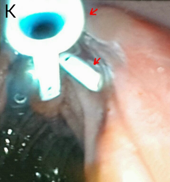
This 26 year old lady had a history of recurrent cholangitis. She had extra hepatic portal venous obstruction with varies at her porta causing compression of the extra hepatic biliary channels. She had previously undergone three ERCPs. The second ERCP showed her dilated left and right biliary systems (A: black arrows) with luminal narrowing of the confluence, CHD & CBD (A: white arrows). Following dilatation with a graduated dilatation catheter, a 10 French plastic stent had been placed in her right system (B: black arrows) but her left system could not be cannulated (B: white arrows). On her 3rd ERCP, the stent had been removed. Now, 4 months later, she presented with cholangitis. Her MRCP images showed the same findings along with stones on the CBD (C: red arrow marks the calculi, black arrows show the narrow CBD, white arrow indicates the narrowing at the hilum and blue arrows show the dilated right and left ductal systems). We booked her for her 4th ERCP. Our guidewire was first manoeuvred into her left system (D: black arrow shows the guidewire and white arrow shows the dilated left system). This was followed by dilatation of the track using a 10 French graduated dilatation catheter (E: white arrows show the dilatation catheter). We then swept the CBD with a biliary balloon (F:black arrows mark the balloon assembly in the CBD) and a  moderate amount of small calculi and sludge was removed (G1: red arrows mark the removed calculi. G2: black arrow shows the "clean sweep" ampulla). We then passed another guidewire in the right system (H: black and white arrows show the two guide wires in the right and left systems, respectively). A 10 French plastic stent of 12 cm length was passed into the right system (I: black arrows mark the stent in the right main duct). A thinner 7 French stent (also of 12 cm length) was then passed into the left system (J: white arrows show the stent in the left main duct while black arrows show the first stent in the right system. K: red arrows show the two stents draining happily in the duodenal lumen).

Saturday, September 20, 2014

No one gets left behind-Retained bile duct stone post cholecystectomy

This 32 year old lady had undergone open cholecystectomy at a peripheral centre. Unfortunately, CBD clearance was incomplete and she was referred to us with a large calculus in the left main duct. She was accompanied by a T-tube cholangiogram (A: Red arrow shows the stone and white arrows shows the T-tube). Before going in, we did a repeat cholangiogram via the T-tube and it showed the stone had migrated to the distal CBD (B: Red arrow shows the meniscus sign, indicating the stone). The T-tube was removed and contrast injection following wire cannulation of the CBD showed that the stone had become lodged in a "recess" of sorts (C: Red arrow), made when the CBD was kinked by the pull of the T-tube. Seeing the considerable size of the stone, we decided to do as wide  a papillotomy as possible (D: White arrow). First weapon of choice was our trusty biliary balloon (E: Black arrow shows the inflated balloon proximal to the stone. Red arrow shows the stone itself. White arrow indicates the marked kink that had developed in the CBD once the T-tube was pulled out). The balloon kept slipping by the side of the stone. Next up was the dormia basket (F: White arrow). The basket was manoeuvred past the stone (G1: Black arrows show the open basket and red arrow marks the stone). The "perp" was finally "apprehended (G2: White arrows show the basket and red arrow shows the captured stone). The stone was pulled out (H: Red arrow shows the stone and black arrows indicate the basket wire around it. I: The retrieved stone seen on a gauze-Homo Sapiens forelimb thumb for size comparison. Oversized glove is evident).

Friday, February 7, 2014

A balloon for every stone

This 50 year old lady had undergone cholecystectomy twenty years earlier. She had now been having recurrent bouts of right hypochondrial pain. An ultrasound scan showed a 12mm stone impacted in the distal CBD. We were greeted by a big fat papilla with the orifice at its lower end (A: white arrow). Multiple attempts to cannulate the CBD were unsuccessful. We decided to do a precut with a needle knife (B). It was a textbook precut and the CBD opening was nicely exposed (C: white arrow shows the CBD opening - somehow appearing suspiciously similar to the Superman insignia). Contrast injection showed two stones, one below the scope and another just above it (D: white arrows). The papillotomy was extended (E) , however the size was insufficient considering the size of the stones. Our next step was to do a sphincteroplasty using an 18mm diameter TTS balloon (F: white arrow shows the waist of the inflated balloon at the ampulla). Following this, two stones were successfully extracted (G1 & G2). A final cholangiogram with an inflated balloon just above the ampulla showed a duct clear of stones (H).

Wednesday, January 29, 2014

Retained CBD stones post cholecystectomy

This lady was referred to us from another hospital. A T tube had been placed following cholecystectomy the post operative ultrasound and T tube cholangiogram suggested that there was a retained stone in the CBD. Contrast injection identified a negative shadow in the upper CBD (A: blue arrow. White arrow marks the T tube). We removed the T tube and performed a balloon sweep which resulted in not one but two stones (B1 & B2: white arrows. The second was a perfectly cut diamond!). An occlusion cholangiogram with contrast injection over a fully inflated balloon (C: white arrow) showed a clear CBD.

Tuesday, January 28, 2014

Tackling a large stone in an old lady

his 80 year old lady had presented with a common bile duct stone (bit unusual in her age group if our past experience was anything to go by). The ampulla was quite swollen (A :white arrow). Contrast injection showed a stone in the upper CBD (B: white arrow). We did a large papillotomy (C: white arrow), keeping in mind the size of the stone. The first attempt at stone extraction was made using a balloon which was maneuvered
across the stone (D: white arrow & E: white arrow shows the balloon inflated behind the stone, marked by the red arrow). The balloon kept slipping past the stone (F: white arrow shows the balloon which has slipped below the stone, marked by the red arrow). We went for the basket next (G: white arrow points to the open basket next to the stone, indicated by red arrow). This was successful and the stone was finally extracted (H1 & H2). Quite large and almost a perfect cylinder (I). Shotgun slugs, anyone?


Second (actually 3rd) ERCP for post transplant biliary leak

This 60 year old gentleman had earlier undergone ERCP and stenting for an anastomotic biliary leakage a few months earlier http://ercp365.bl...