Showing posts with label juxtapapillary diverticulum. Show all posts

Friday, April 25, 2014

Extra large diverticulae flanking ampulla

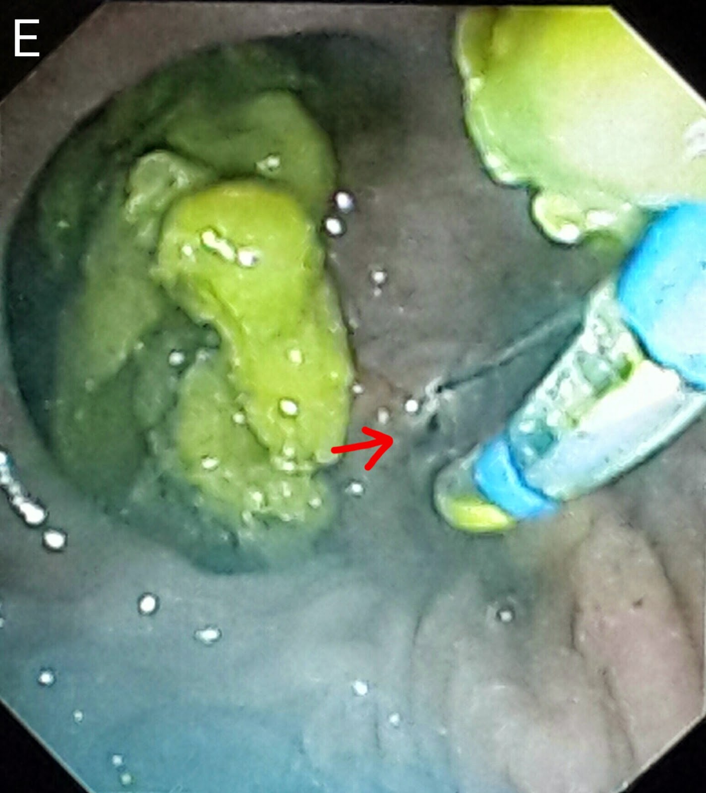
This 60 year old gentleman had been referred following the development of classical obstructive jaundice signs. His ultrasound reported an ampullary mass lesion. The CT report showed a greatly distended CBD with a large calculus at the terminal end (A: white arrow posts to the stone and black arrows delineate the CBD). There was a "whoa" moment the moment the papilla came into view as it was flanked by two huge diverticulae stuffed with food (B: red arrows. White arrow points to the papilla). The scope kept slipping and cannulating the ampulla was trickier than expected (C). Contrast injection showed a dilated CBD with a large stone near the lower end (D: black arrow). We did a small sphincterotomy (E: red arrow). A 10 French plastic stent of 10 cm length was then deployed (F1: red arrow shows the stent and blue arrow points to the stent assembly. F2: black arrows show the stent). The patient was referred to the surgeon for cholecystectomy and bile duct clearance. 

Wednesday, April 9, 2014

The brothers diverticulae

This 35 year old lady had been referred after a history of biliary type pain and an ultrasound which reported gallbladder sludge and a dilated CBD with a distal stricture. The MRCP report confirmed the ultrasound findings of CBD dilatation (A: red arrows) and a distal stricture (A: white arrow). Going in,  we were greeted with two diverticulae (B: red arrows) sitting on top of the papilla (B: white arrow). Contrast injection showed a picture similar to the MRCP of a dilated bile duct (C: red arrows) with distal narrowing (C: white arrow). We did a papillotomy which was limited by the close proximity of the lower edge of the diverticulae (D: red arrow). The CBD was swept with a biliary balloon (E: white arrow). The limited papillotomy necessitated a sphincteroplasty. We used a TTS balloon of 15-18mm expansion range (F1 & F2) and dilated the papilla to 15 mm (G1 & G2 showing the balloon expanding at the papilla) which was maintained for 30 seconds (G3: black arrows mark the balloon. Red arrow shows the waist forming at the papilla). The end result was a wide open papilla (H). 


Saturday, February 8, 2014

Diverticula Galore!

This 84 year old lady had been admitted with cholangitis. Her ultrasound scan indicated a dilated CBD with a distal stricture. Her CA 19-9 level was also normal. The MRCP film confirmed the ultrasound findings of a dilated CBD with a distal narrowing (A: white arrow). Starting our ERCP, we were greeted with a duodenum full of diverticulae. Fortunately, the papilla was not at the bottom of one of these (B: white arrows indicating two adjacent diverticulae, with food residue in the left one. Blue arrow indicating the papilla. Black arrow indicating the duodenal lumen). Thankfully it didn't take too long to cannulate the papilla (C). Contrast injection showed the same findings as the MRCP (D) and we suspect that the lower narrowing in the CBD could be attributed to the diverticulae. We passed a 10 French 7cm long plastic stent (E1 & E2: white arrows).

Second (actually 3rd) ERCP for post transplant biliary leak

This 60 year old gentleman had earlier undergone ERCP and stenting for an anastomotic biliary leakage a few months earlier http://ercp365.bl...