Showing posts with label post cholecystectomy bile leak. Show all posts

Tuesday, May 20, 2014

Follow up case of post cholecystectomy biliary leak

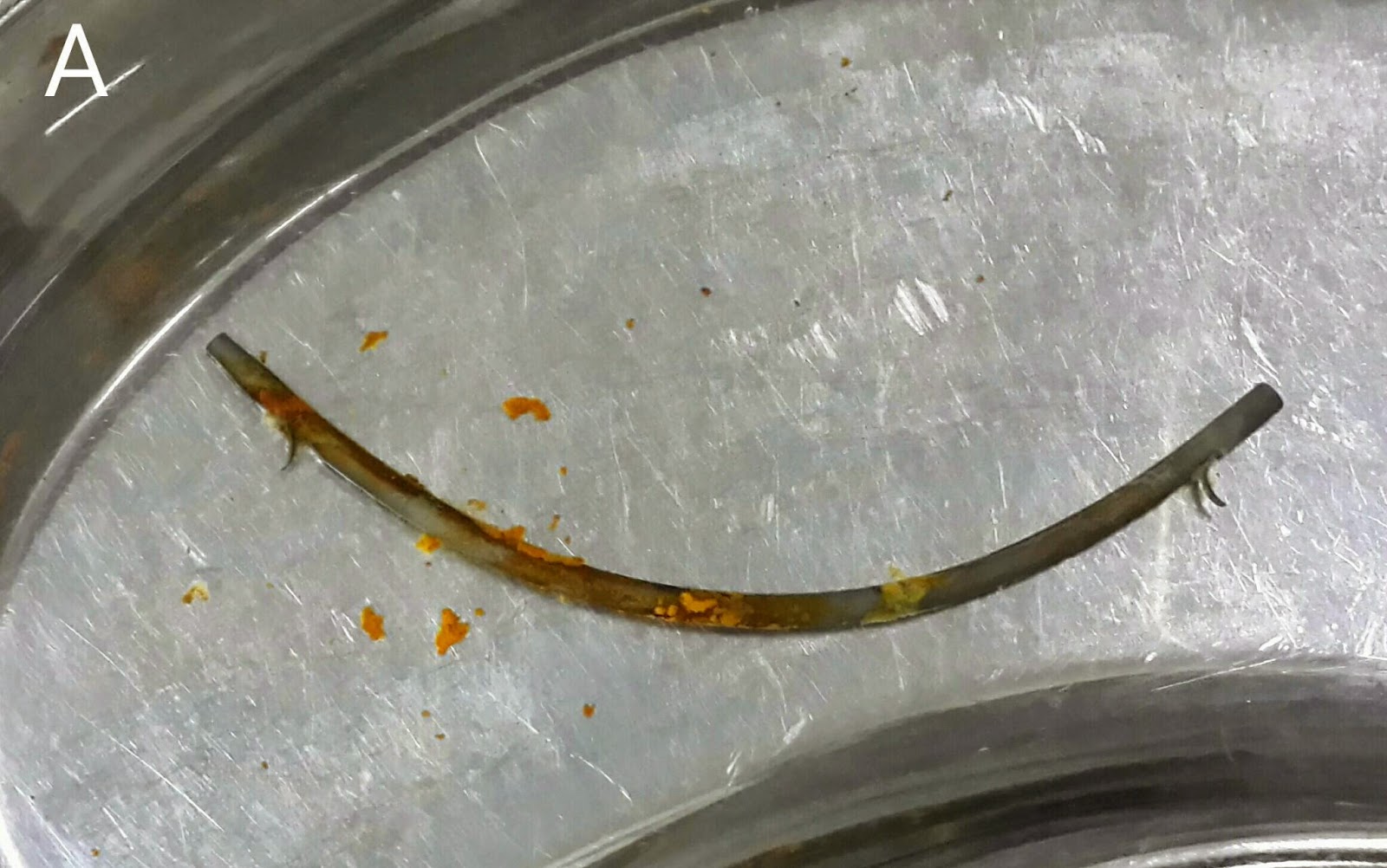
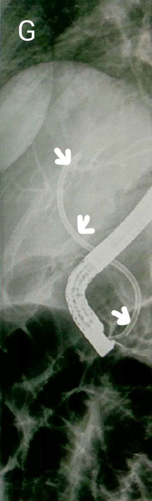
This 38 year old lady had previously undergone biliary stunting for a bile leak http://ercp365.blogspot.com/2013/12/and-another-bile-leak-following.html which had occurred following cholecystectomy. We had placed a 10 cm long 10 French plastic stent. We removed the stent (A). The sludge coating prompted us to do a biliary sweep with a stone extraction balloon (B: white arrow shows the papilla with a previous sphincterotomy and the balloon assembly being inserted). The CBD was swept (C: black arrows shows the inflated Ballon in the CBD). Gan occlusion cholangiogram with the balloon inflated at the distal end of the CBD was done (D: black arrow shows the inflated balloon. White arrows delineate the CBD with no leakage). The patient was discharged. 

Friday, May 9, 2014

Major biliary tree disruption post surgery

This 52 year old lady had been referred from another hospital following bile duct injury during open cholecystectomy. She underwent a repeat laparotomy after her gall bladder surgery for the leak. A drain was placed in the sub hepatic space and the patient was subsequently sent to us. Her MRCP wasn't the best of images we had seen, with major motion artifacts (problem with holding her breath, we suppose). A disruption in the bile duct (A: red arrow) and leakage (A: blue arrow) was clear. Contrast injection after CBD cannulation showed the extent of damage, with structures, leaks and what appeared to be an uncommon  anatomical variant of the biliary tree (B: green arrows show the leak. Blue arrow shows a major stricture. Black & white arrows show what appear to be right and left hepatic ducts but without the classical confluence morphology we're used to seeing. Red arrow indicates a 0.035 guidewire which has been placed into the right system). The right system,where we had managed to place the wire, was dilated with a 10 French graduated dilatation catheter (C: black arrows). We then left one wire in the right duct and made multiple attempts to place the second wire into the left duct in order to dilate it (D1 to D4: black arrow shows the first wire in the right system and white arrow shows the second wire as we attempt to place it in the left duct). Alas we were unsuccessful. A decision was made to stent the right duct. A stent assembly was then passed. As we passed the stent over it, the force required to pass the strictured segment (despite dilatation done earlier) caused the stent assembly to warp into the area of leakage (E: white arrows show the stent assembly and black arrow shows the assembly bent into the leak area). A 10 French stent of 12 cm length was then deployed (F1 &  F2: white arrows). 


Friday, April 25, 2014

Lost to follow up

This 45 year old gentleman had suffered from bile duct injury whilst undergoing an open cholecystectomy 10 months ago. The surgeons placed a drain (A: red arrow shows the drain. White arrow shows the large leak-almost looking like the gallbladder in shape. Blue arrow shows the main bile duct). An MRI was done soon after and confirmed the leakage (B: red arrows show the bile leak). He was reffered to our facility and we had placed a 10 French 10cm long plastic stents to bridge the leak. (C: white arrows show the stent) . This was about ten months ago. He was then lost to follow up. He resurfaced at our outpatient out of curiosity asking what was to be done about the Stent we had placed. When questioned as to why he hadn't turned up a month after the procedure, he said he had been told by his physician that his condition has been cured and he needs no more treatment. The moment we went in for ERCP, we were greeted by a normal ampulla with no sign of his placed stent. We thought it had migrated inwards but that wasn't the case as it didn't show up on flouroscopy either. One thing we noted was almost absent bike flow from the ampulla and normal flow from another opening just above it (D: Blue arrow marks the ampulla and white arrow indicates the second opening above it with smeared bile). This second opening was obviously a fistula. Cannulation and contrast injection showed no bile leakage (E: white arrows show the bile duct. Even the fistulous track wasn't outlined). We concluded that the Stent had slipped out and his bole was driving through the fistula. The procedure was ended by a sphincterotomy which was extended to include the fistulous opening above the ampulla (F: white arrow). 


Wednesday, April 2, 2014

Techniques to pull an old friend out of trouble

This 34 year old lady had been referred to our centre two years ago with retained CBD stones and bike duct injury following cholecystectomy.  She had a T tube placed. Her old ERCP films showed small stones (A: white arrows) and the T tube (A: red arrows ). The T tube was removed and the CBD was swept with balloon (B:white arrow shows the inflated balloon in the CBD). We placed a 10 French plastic sent of 12 cm length (C: white arrows). She was sent back to her referring centre. We had hoped to see her again after 8 weeks to remove the stent but she was lost to follow up. Fast forward two years and she expectedly developed biliary pain,  jaundice and fever. She went back to her original hospital where a diagnosis of blocked stent and biliary stones was made and she was sent to us. We saw our old stent which had extruded quite a bit forward into he duodenum (D: red arrow) and we couldn't properly place a snare to grab it. Initially we tried to pull it out using an inflated balloon placed adjacent to it in the CBD (E: blue arrow shows the stent and white arrow indicates the balloon adjacent to it). Our next move was to use a large bipod forceps (F1) to grab the stent (F2) and slide it back in the papilla (F3). The stent was now easily grabbed with snare (G: white arrow) and pulled out (H). Repeat contrast injection in the CBD showed a lot of stones (I: black arrows). A papillotomy was done (J). The routine technique if sweeping the lower CBD to gauge adequacy of papillotomy resulted in a lot of sludge being removed (K1 &  K2). The CBD was the swept with balloon (L: black arrow shows the inflated balloon in the upper CBD) which resulted in a lot of stones (M) and sludge (N)  being removed. The final cholangiogram showed a CBD clear of stones (O: red arrows). 

Thursday, January 2, 2014

Bile duct injury and calculi post cholecystectomy

This 35 year old lady had undergone a laparosocpic cholecystectomy at another hospital three months ago. She had been re-admitted with an acute abdomen and the surgeons reported a rent in the CBD and placed a T-tube. She was then referred to us for CBD stenting. Her T-tube cholangiogram showed leakage of contrast (A: white arrow) and what appeared to be a stone (A: red arrow). Contrast injection on ERCP confirmed the cholangiogram findings and showed both the leak and the stone (B: white and red arrows respectively). Curiously, the T-tube was not outlined and we Suspected that it may have gotten dislodged. We did a wide papillotomy (C) and swept the CBD with a stone extraction balloon (D) which yielded a stone (E: white arrow). An occlusion cholangiogram an inflated balloon (F: white arrow) showed a clear CBD. We ended the procedure by placing a 12 cm long single pigtail stent of 10 French diameter (G: white arrows). Single pigtail stent being necessary to anchor the stent inside the CBD keeping in mind the wide papillotomy that had been done. 

Monday, December 30, 2013

Bike leak following laparosocpic cholecystectomy

This 54 year old gentleman had been referred to our unit with bile leak following a laparosocpic cholecystectomy. His MRCP showed the leak and collection (A: white arrow). Contrast injection on ERCP confirmed the MRCP report and the leakage was clearly seen (B1 & B2: white arrows). A 10cm long 10 French plastic stent was passed to bridge the leak (C: white arrows show the stent. Red arrow marks the leaked contrast).

Wednesday, December 11, 2013

And another bile leak following cholecystectomy

This 38 year old lady had the same problem as the previous case: A cholecystectomy gone wrong. Her MRCP showed a small leak (A: white arrow. Green arrow shows the pancreatic duct). On ERCP, the same leak was outlined on contrast injection (B and C: white arrows).   A 10 cm long plastic stent of 10 French diameter was placed (D: white arrows).

Stenting a biliary leak

A 35 year old lady had been referred to our center after a bile duct injury during cholecystectomy. A drain and been placed in the sub hepatic space. On ERCP, the placed drainage tube (A: blue arrow) was seen just below the level of confluence (A: yellow arrow). The negative shadows seen in the image are actually air bubbles (A: green arrow). Interestingly, the contrast injected didn't show any leakage per se and went straight into the drainage tube instead. We concluded that the tube must be abutting the biliary tree. A 12 cm 10 French plastic stent was placed to bridge the site of leakage (B: blue arrows).

Sunday, September 15, 2013

Biliary stent migrated into CBD

This lady had a bile duct stent placed after a post cholecystectomy biliary leak. We had gone in to retrieve the stent. It had unexpectedly migrated into the CBD. Using the traditional snare to grab it was not possible. So we tried a dormia basket first (A: arrows showing the extent of the open basket), which didn't work. We tried a Soehendra stent retriever but couldn't screw the stent. We lastly tried a papillotomy to see if we could further expose the stent. That too didn't help. Finally grabbing the exposed bit by forceps was tried (B: green arrow showing the stent. Red arrow points to the forceps). Again, unsuccessful. We'll reattempt in a couple of weeks.

Second (actually 3rd) ERCP for post transplant biliary leak

This 60 year old gentleman had earlier undergone ERCP and stenting for an anastomotic biliary leakage a few months earlier http://ercp365.bl...