Showing posts with label dormia basket. Show all posts

Saturday, September 20, 2014

No one gets left behind-Retained bile duct stone post cholecystectomy

This 32 year old lady had undergone open cholecystectomy at a peripheral centre. Unfortunately, CBD clearance was incomplete and she was referred to us with a large calculus in the left main duct. She was accompanied by a T-tube cholangiogram (A: Red arrow shows the stone and white arrows shows the T-tube). Before going in, we did a repeat cholangiogram via the T-tube and it showed the stone had migrated to the distal CBD (B: Red arrow shows the meniscus sign, indicating the stone). The T-tube was removed and contrast injection following wire cannulation of the CBD showed that the stone had become lodged in a "recess" of sorts (C: Red arrow), made when the CBD was kinked by the pull of the T-tube. Seeing the considerable size of the stone, we decided to do as wide  a papillotomy as possible (D: White arrow). First weapon of choice was our trusty biliary balloon (E: Black arrow shows the inflated balloon proximal to the stone. Red arrow shows the stone itself. White arrow indicates the marked kink that had developed in the CBD once the T-tube was pulled out). The balloon kept slipping by the side of the stone. Next up was the dormia basket (F: White arrow). The basket was manoeuvred past the stone (G1: Black arrows show the open basket and red arrow marks the stone). The "perp" was finally "apprehended (G2: White arrows show the basket and red arrow shows the captured stone). The stone was pulled out (H: Red arrow shows the stone and black arrows indicate the basket wire around it. I: The retrieved stone seen on a gauze-Homo Sapiens forelimb thumb for size comparison. Oversized glove is evident).

Tuesday, January 28, 2014

Tackling a large stone in an old lady

his 80 year old lady had presented with a common bile duct stone (bit unusual in her age group if our past experience was anything to go by). The ampulla was quite swollen (A :white arrow). Contrast injection showed a stone in the upper CBD (B: white arrow). We did a large papillotomy (C: white arrow), keeping in mind the size of the stone. The first attempt at stone extraction was made using a balloon which was maneuvered
across the stone (D: white arrow & E: white arrow shows the balloon inflated behind the stone, marked by the red arrow). The balloon kept slipping past the stone (F: white arrow shows the balloon which has slipped below the stone, marked by the red arrow). We went for the basket next (G: white arrow points to the open basket next to the stone, indicated by red arrow). This was successful and the stone was finally extracted (H1 & H2). Quite large and almost a perfect cylinder (I). Shotgun slugs, anyone?


Sunday, October 27, 2013

The stone that wouldn't come out

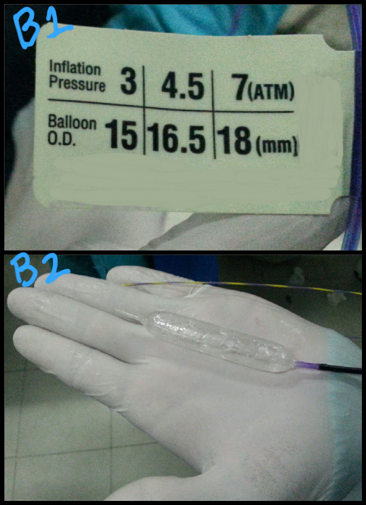
This 28 year old gentleman had what appeared to be "a soft mass with acoustic shadow" in his distal CBD. Contrast injection on ERCP showed a rounded stone in the mid CBD (A: green arrow). After a wide papillotomy, we attempted to extract the stone with an extraction balloon but were unsuccessful. We decided to do a sphincteroplasty with a wire guided balloon (B1 & B2: the balloon specifications and inflation check prior to insertion. C1 to C4: the balloon being placed into the papilla and inflated. C5: green arrows show the extent of dilated balloon on fluoroscopy). Even after sphincteroplasty, the stone could not be removed with the extraction balloon and we could clearly see it stuck in the CBD through the wide papillotomy (D: green arrow). We then decided to use the extraction basket to remove the lodged stone (E: white arrows show the open basket in the CBD). This was rewarded with success and the stone was removed (F: green arrow shows the stone, smeared with blood, lying in the duodenal lumen). 

Saturday, October 26, 2013

Multiple CBD stones requiring different removal techniques

This 40 year old lady had a pre ERCP ultrasound which indicated a single 9mm stone in the distal CBD. Going in, we were encountered by a swollen ampulla (A: green arrow), suggesting an impacted stone. Dye injection showed a grossly dilated CBD (B: green arrows) and negative shadows (B :red arrows). The stones could not be removed even after a wide papillotomy. So we went for a sphincteroplasty with an 18mm TTS (through the scope) wire guided balloon (C1 & C2 and D: green arrow showing the inflated balloon at the ampulla). One stone was retrieved (E: green arrow). Repeat cholangiogram showed more stones in the mid CBD (F: green arrow). The remain stones were removed with an extraction basket (G1: green arrow marks the stone and red arrow indicates the basket wires. G2: red arrow marks the stone trapped in the basket wires)

Sunday, September 15, 2013

Biliary stent migrated into CBD

This lady had a bile duct stent placed after a post cholecystectomy biliary leak. We had gone in to retrieve the stent. It had unexpectedly migrated into the CBD. Using the traditional snare to grab it was not possible. So we tried a dormia basket first (A: arrows showing the extent of the open basket), which didn't work. We tried a Soehendra stent retriever but couldn't screw the stent. We lastly tried a papillotomy to see if we could further expose the stent. That too didn't help. Finally grabbing the exposed bit by forceps was tried (B: green arrow showing the stent. Red arrow points to the forceps). Again, unsuccessful. We'll reattempt in a couple of weeks.

Second (actually 3rd) ERCP for post transplant biliary leak

This 60 year old gentleman had earlier undergone ERCP and stenting for an anastomotic biliary leakage a few months earlier http://ercp365.bl...