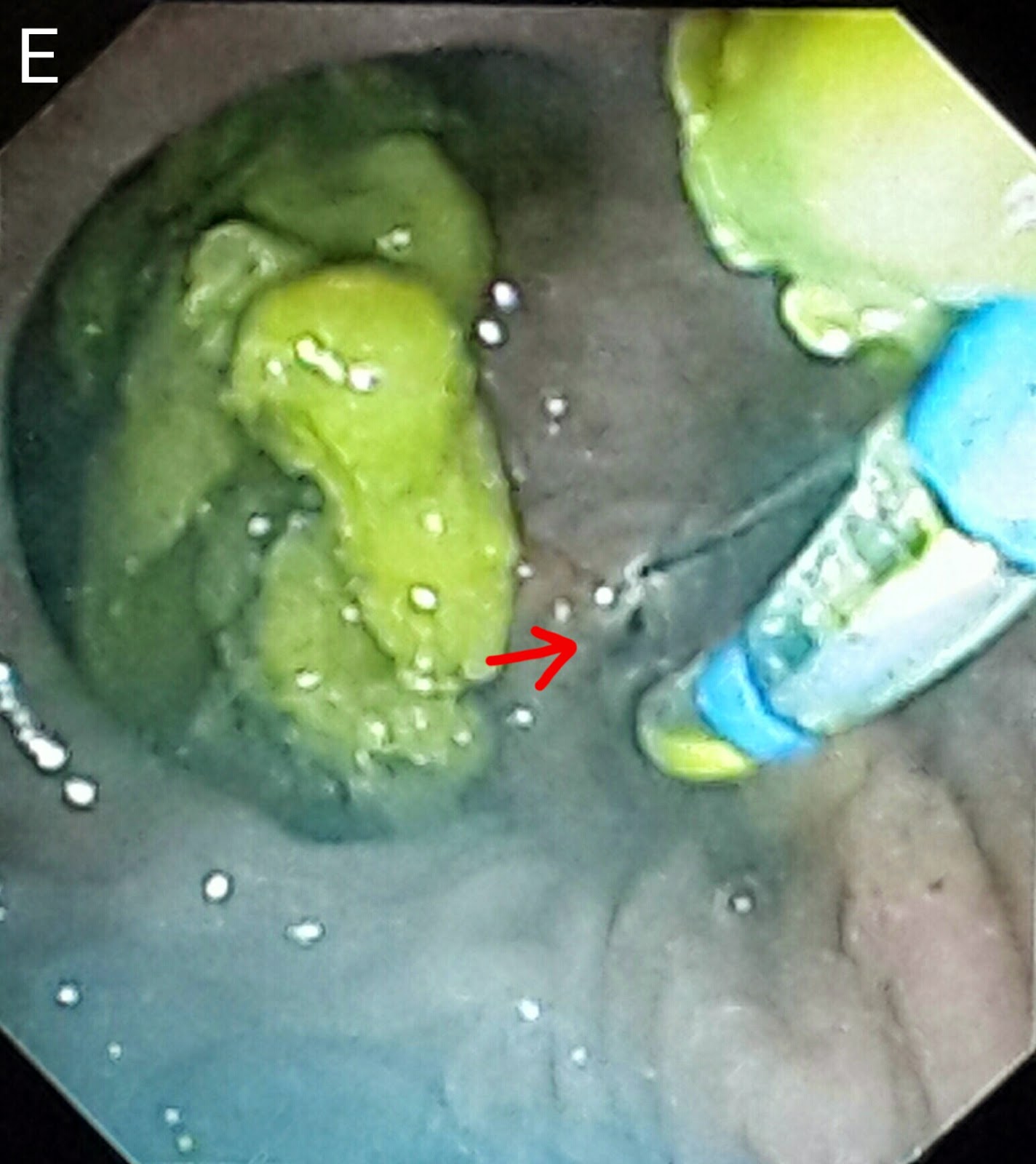
This 60 year old gentleman had been referred following the development of classical obstructive jaundice signs. His ultrasound reported an ampullary mass lesion. The CT report showed a greatly distended CBD with a large calculus at the terminal end (A: white arrow posts to the stone and black arrows delineate the CBD). There was a "whoa" moment the moment the papilla came into view as it was flanked by two huge diverticulae stuffed with food (B: red arrows. White arrow points to the papilla). The scope kept slipping and cannulating the ampulla was trickier than expected (C). Contrast injection showed a dilated CBD with a large stone near the lower end (D: black arrow). We did a small sphincterotomy (E: red arrow). A 10 French plastic stent of 10 cm length was then deployed (F1: red arrow shows the stent and blue arrow points to the stent assembly. F2: black arrows show the stent). The patient was referred to the surgeon for cholecystectomy and bile duct clearance.