Showing posts with label stent removal. Show all posts

Tuesday, May 20, 2014

Follow up case of post cholecystectomy biliary leak

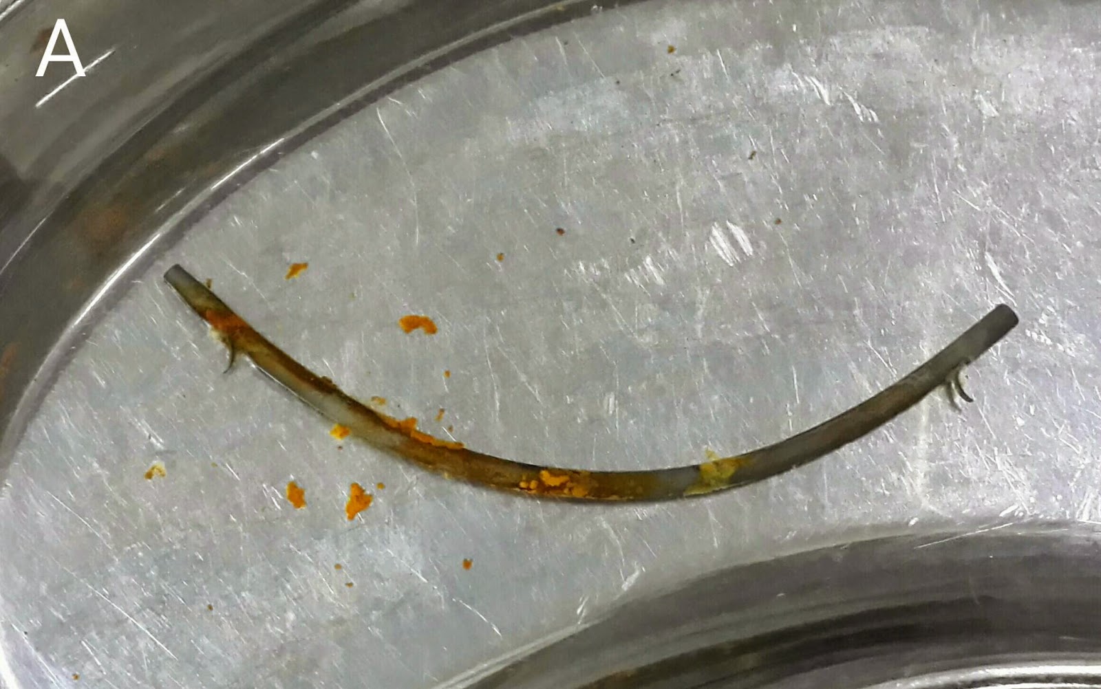
This 38 year old lady had previously undergone biliary stunting for a bile leak http://ercp365.blogspot.com/2013/12/and-another-bile-leak-following.html which had occurred following cholecystectomy. We had placed a 10 cm long 10 French plastic stent. We removed the stent (A). The sludge coating prompted us to do a biliary sweep with a stone extraction balloon (B: white arrow shows the papilla with a previous sphincterotomy and the balloon assembly being inserted). The CBD was swept (C: black arrows shows the inflated Ballon in the CBD). Gan occlusion cholangiogram with the balloon inflated at the distal end of the CBD was done (D: black arrow shows the inflated balloon. White arrows delineate the CBD with no leakage). The patient was discharged. 
Tuesday, February 25, 2014

A blocked stent within a blocked stent

This 78 year old gentleman had undergone ERCP for a Klastkin tumor three months ago. The procedure was unsuccessful and he was then referred for a PTBS (percutaneous transhepatic biliary stenting) following which a self expanding metallic stent was placed. Two months later, his stent was blocked and he underwent an ERCP in which a double pigtail 10 French diameter stent of 10 cm length was placed within the metallic stent (A: white arrows show the extent of the metallic stent and black arrows indicate the curved ends of the plastic stent within it ). This plastic stent lasted for over a month before being blocked (B: white arrow marks the blocked metal stent and blue arrows indicate the curled distal end of the plastic stent). The plastic stent was retrieved with a snare (C). Contrast injection outlined the intraheptic channels and the guidewire was advanced beyond the stent (D: white arrow shows the extent of the metallic PTBS stent and black arrow shows the guidewire beyond its upper end ). On retrieval, the sheath of the guidewire tip was broken and left inside (E: black arrow). The next image shows a more clear outlining of the left ductal system (F: white arrows). The metal stent and biliary tree was flushed with saline and an 11 cm straight plastic stent of 10 French diameter was placed (G: white arrows show the metal stent and black arrows show the new plastic stent protruding at both ends. H: the new plastic stent within the metal stent as seen in the duodenum).

Sunday, February 2, 2014

Third follow-up ERCP for post transplant anastomotic biliary stricture

This 37 year old hospital nurse had undergone living donor liver transplant for Hepatitis C related liver disease. She had developed an anastomotic biliary stricture for which we had done two ERCPs previously, one in September http://ercp365.blogspot.com/2013/09/post-liver-transplant-anastomotic_10.html (single 12 French plastic stent passed) and second in December (Two 10 French plastic stents placed). The latest ultrasound suggested blocked stents. This was confirmed visually (A). Both the stents were removed with snare (B). Contrast injection showed the narrowing in the anastomotic site (C: white arrow). We decided to use a 3cm long, 10mm diameter TTS balloon rated at 90 psi (D) for stricturoplasty. The balloon was inflated in the stricture area (E1 to E3: white arrows). We then swept the CBD with a balloon to clear any sludge. An occlusion cholangiogram showed successful treatment of the stricture (F: white arrow). Although we were thinking pre procedure of placing three stents, the stricturoplasty results made us decide against it. No stents were placed and the patient will now be followed with serial bilirubin levels and ultrasound scans.

Monday, December 30, 2013

Blockage of biliary stent placed for post transplant anastomotic stricture

This gentleman had undergone a living donor liver transplant approximately a month ago. A plastic biliary stent had been placed for an anastomotic stricture about three weeks after transplant. This was eight days ago. His total bilirubin level dropped from 15 mg/dL to 10.5 mg/dL in the 72 hours following stent placement. He then experienced fever along with rising bilirubin levels. We expected that the stent was blocked ( we had noted a significant amount of sludge along with bile flowing from the stent the first time we had placed it). Sure enough, we were met with a choked stent on ERCP (A1). We grabbed it with a snare (A2 & A3) and removed it (A4). We then generously flushed the bile duct with saline to wash out all debris (B1). A small papillotomy was also done to assist in drainage of the sludge (B2). Repeat contrast injection was done to check the status of the stricture. Sure enough, the stricture was present just behind the endoscope (C: white arrow). Since it had been only four weeks since the transplant, we avoided balloon dilatation of the strictured area (We had already "stretched" protocol by placing stent larger than 7 French diameter just three weeks after transplant in this patient. We had placed a 10 French stent and vendors that had been blocked). A similar 10 French plastic stent of 10 cm length was placed (D1 &  D2: white arrows). A gush of bile and sludge was seen (E). We're keeping our fingers crossed as there is a probability that this stent might become blocked as well over the next one to two weeks.

Tuesday, September 24, 2013

Post cholecystectomy CBD stricture

This 40 year old lady had presented with a stricture of the CBD at the level of the cystic duct following a cholecystectomy. We had passed a single plastic stent of 10F diameter two months ago. She currently had normal bilirubin levels. This was a follow up ERCP. We removed the previously placed stent with a snare. Dye injection showed a CBD stricture at the level of cystic duct (A: green arrow). We had planned to place two new 10F x 12 cm plastic stents. The first stent was placed across the stricture (B: green arrow). The guidewire was  then manoeuvred along the first stent (C: green arrow marks the guidewire red arrows mark the first stent). Unfortunately, the sheath on the tip of the guidewire broke off and couldn't be retrieved (D: red arrow). A new guidewire was then passed along the same route and the second stent was placed parallel to the first one ( E1 & E2: green arrows & F).

Wednesday, August 28, 2013

Biliary stent penetrating opposite duodenal wall

Not a common sight. We had gone for stent removal and attempt stone extraction (the patient had previously had cholangitis and stone extraction was incomplete. A plastic stent had been placed). The stent had penetrated the opposite duodenal wall, a closed perforation. We had to wrestle a bit with the forceps, pushing the stent back into the CBD. It was then removed with a snare.

Second (actually 3rd) ERCP for post transplant biliary leak

This 60 year old gentleman had earlier undergone ERCP and stenting for an anastomotic biliary leakage a few months earlier http://ercp365.bl...