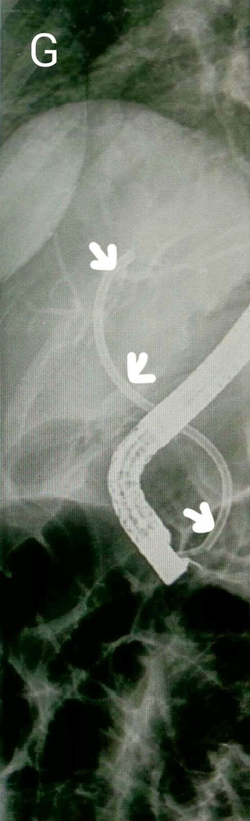
This 35 year old lady had undergone a laparosocpic cholecystectomy at another hospital three months ago. She had been re-admitted with an acute abdomen and the surgeons reported a rent in the CBD and placed a T-tube. She was then referred to us for CBD stenting. Her T-tube cholangiogram showed leakage of contrast (A: white arrow) and what appeared to be a stone (A: red arrow). Contrast injection on ERCP confirmed the cholangiogram findings and showed both the leak and the stone (B: white and red arrows respectively). Curiously, the T-tube was not outlined and we Suspected that it may have gotten dislodged. We did a wide papillotomy (C) and swept the CBD with a stone extraction balloon (D) which yielded a stone (E: white arrow). An occlusion cholangiogram an inflated balloon (F: white arrow) showed a clear CBD. We ended the procedure by placing a 12 cm long single pigtail stent of 10 French diameter (G: white arrows). Single pigtail stent being necessary to anchor the stent inside the CBD keeping in mind the wide papillotomy that had been done.