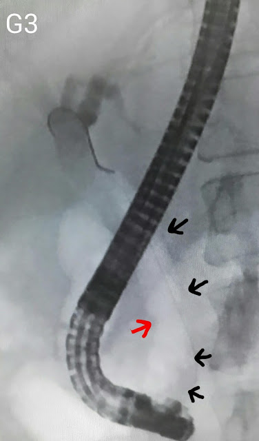
This 35 year old lady had been referred after a history of biliary type pain and an ultrasound which reported gallbladder sludge and a dilated CBD with a distal stricture. The MRCP report confirmed the ultrasound findings of CBD dilatation (A: red arrows) and a distal stricture (A: white arrow). Going in, we were greeted with two diverticulae (B: red arrows) sitting on top of the papilla (B: white arrow). Contrast injection showed a picture similar to the MRCP of a dilated bile duct (C: red arrows) with distal narrowing (C: white arrow). We did a papillotomy which was limited by the close proximity of the lower edge of the diverticulae (D: red arrow). The CBD was swept with a biliary balloon (E: white arrow). The limited papillotomy necessitated a sphincteroplasty. We used a TTS balloon of 15-18mm expansion range (F1 & F2) and dilated the papilla to 15 mm (G1 & G2 showing the balloon expanding at the papilla) which was maintained for 30 seconds (G3: black arrows mark the balloon. Red arrow shows the waist forming at the papilla). The end result was a wide open papilla (H).
I am a gastroenterologist. This is a blog of the ERCPs and related endoscopic procedures carried out at my department. Dr Adnan Salim.
Subscribe to:
Post Comments (Atom)
Second (actually 3rd) ERCP for post transplant biliary leak
This 60 year old gentleman had earlier undergone ERCP and stenting for an anastomotic biliary leakage a few months earlier http://ercp365.bl...

-
This 22 year old lady had undergone living donor liver transplant at our centre for hepatitis B related liver disease. She developed an ana...
-
This 25 year old gentleman had undergone a previous ERCP for an anastomotic biliary stricture. http://ercp365.blogspot.com/2015/04/post-li...
-
A healthy 48 year old gentleman with a recent history of jaundice. The MRCP outlined a dilated proximal CBD with a small portion of the dist...


No comments:
Post a Comment