Showing posts with label sphincteroplasty. Show all posts

Friday, July 10, 2015

Retrieving a retained stone post cholecystectomy

This 35 year old lady had undergone cholecystectomy at a private facility last month which was complicated by a biliary leak. She was reopened and a T-tube was  placed. A post procedure T-tube cholangiogram showed a calculus in the CBD (A: Red arrow). The T-tube was then removed and she was referred to our centre. An MRCP was done which confirmed the cholangiogram findings of a CBD stone (B: White arrow). Contrast injection on ERCP showed a vague negative shadow in the upper CBD (C: Blue arrow indicates the concerned area while white arrow shows an inflated biliary balloon at the distal CBD). Sphinchteroplasty was planned there wasn't margin for a full blooded sphincterotomy and a 12-15mm TTS balloon rated at 3-8 ATM (D) was used to dilate the ampulla (E: Black arrow shows the inflated balloon). The patient experienced pain despite high dose IV analgesics so we restricted ourselves to 12mm dilatation for 10 seconds (F: Black arrow shows the post sphincteroplasty ampulla). At this point we were skeptical as to whether the stone would be retrieved. Nevertheless a sweep was made and successful stone extraction was done (G1 & G2: Black arrows show the stone being retrieved). An occlusion cholangiogram with an inflated ballon at the ampulla (H: White arrow) showed a clear CBD. 
5


Sunday, October 27, 2013

The stone that wouldn't come out

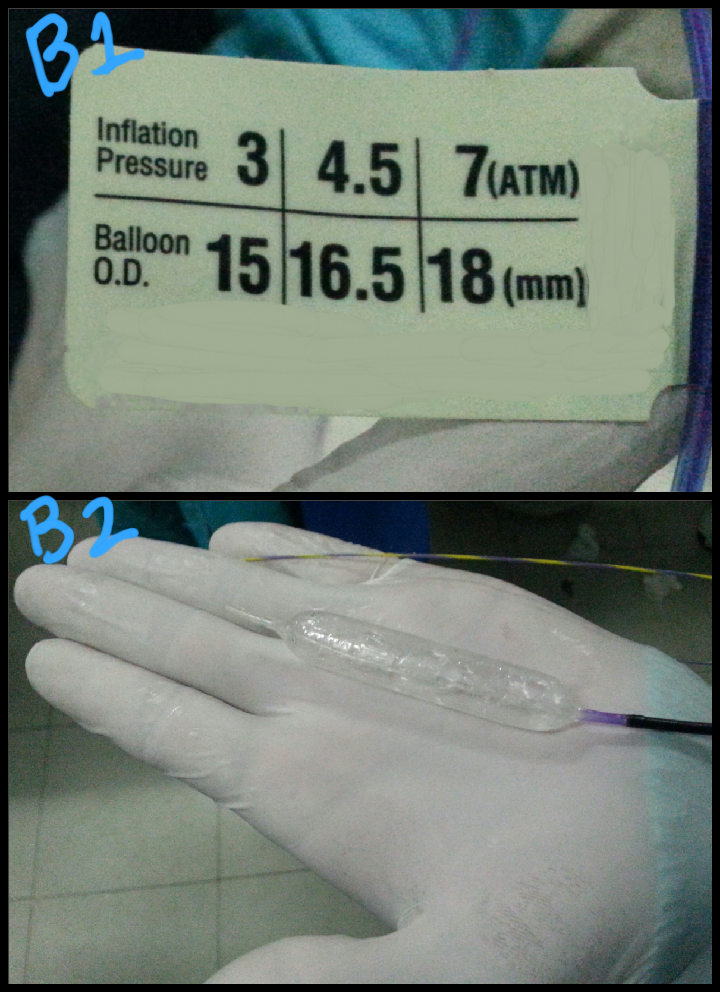
This 28 year old gentleman had what appeared to be "a soft mass with acoustic shadow" in his distal CBD. Contrast injection on ERCP showed a rounded stone in the mid CBD (A: green arrow). After a wide papillotomy, we attempted to extract the stone with an extraction balloon but were unsuccessful. We decided to do a sphincteroplasty with a wire guided balloon (B1 & B2: the balloon specifications and inflation check prior to insertion. C1 to C4: the balloon being placed into the papilla and inflated. C5: green arrows show the extent of dilated balloon on fluoroscopy). Even after sphincteroplasty, the stone could not be removed with the extraction balloon and we could clearly see it stuck in the CBD through the wide papillotomy (D: green arrow). We then decided to use the extraction basket to remove the lodged stone (E: white arrows show the open basket in the CBD). This was rewarded with success and the stone was removed (F: green arrow shows the stone, smeared with blood, lying in the duodenal lumen). 

Saturday, October 26, 2013

Multiple CBD stones requiring different removal techniques

This 40 year old lady had a pre ERCP ultrasound which indicated a single 9mm stone in the distal CBD. Going in, we were encountered by a swollen ampulla (A: green arrow), suggesting an impacted stone. Dye injection showed a grossly dilated CBD (B: green arrows) and negative shadows (B :red arrows). The stones could not be removed even after a wide papillotomy. So we went for a sphincteroplasty with an 18mm TTS (through the scope) wire guided balloon (C1 & C2 and D: green arrow showing the inflated balloon at the ampulla). One stone was retrieved (E: green arrow). Repeat cholangiogram showed more stones in the mid CBD (F: green arrow). The remain stones were removed with an extraction basket (G1: green arrow marks the stone and red arrow indicates the basket wires. G2: red arrow marks the stone trapped in the basket wires)

Second (actually 3rd) ERCP for post transplant biliary leak

This 60 year old gentleman had earlier undergone ERCP and stenting for an anastomotic biliary leakage a few months earlier http://ercp365.bl...