Showing posts with label biliary balloon. Show all posts

Thursday, April 16, 2015

Second ERCP for post transplant anastomotic biliary stricture (CASE J)

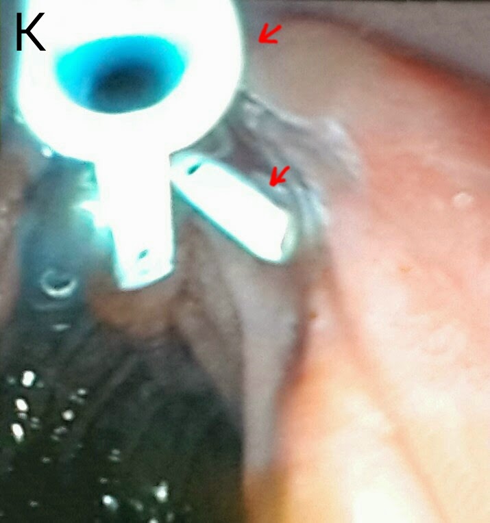
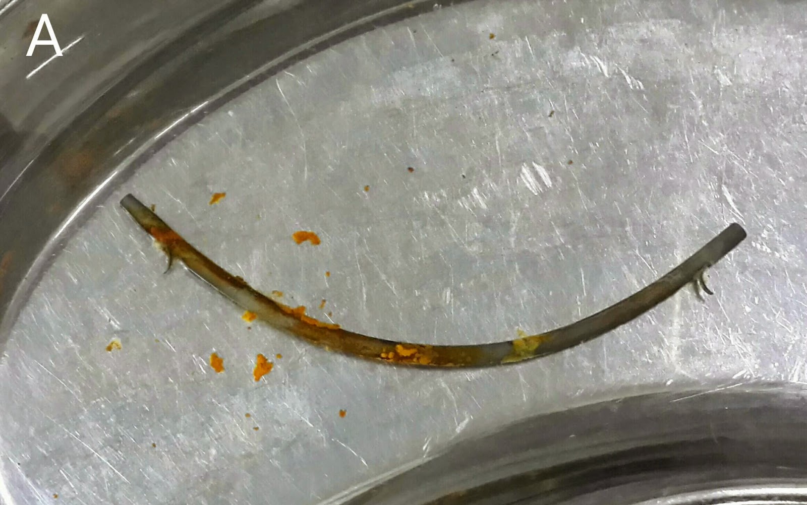
This 56 year old gentleman underwent a living related transplant for liver cirrhosis secondary to Hepatitis C around March 2014. He had his first ERCP in December 2014 (an earlier attempt in October 2014 had been unsuccessful). Contrast injection identified a sharp L-shaped angulation of the left hepatic duct, crossing in front of the right duct (A: white arrows). The right duct was seen behind it (A: black arrow). A 7 French stent of 12cm length was deployed in the left duct after dilating the track with a nominal 7 French graduated dilatation catheter. He again developed burning of the soles of his feet and pruritis along with rising ALP. We decided to remove his stent. On ERCP, it was seen to have migrated inside the CBD (B: Black arrows). We retrieved it using a biliary balloon inflated next to it (C: black arrows show the stent and white arrow marks the balloon adjacent to the stent. D: a single balloon sweep had resulted in some pullout and straightening of the proximal sharply bent portion of the stent-compare with C. E: white arrow shows the retrieved stent). Contrast injection post stent retrieval resulted in clear outlining of the right duct (F: black arrow) however the left L-shaped duct was only marginally outlined (F: white arrow). A balloon assembly was positioned in the right ductal system (G: black arrow) which was then inflated with full obliteration of the waist (H: black arrow). Next we manoeuvred our guidewire into the L-shaped left duct (I: black arrow). Pushing the papillotome over the wire and injection of contrast now outlined the left duct in its sharply bent form (J: black arrows. Compare with A). A 7 French double pigtail stent of 12 cm length was deployed (K: black arrows. Note the marked coil in the proximal portion of the stent. Not quite the desired result but we'll keep an eye on him nevertheless to see how it drains). A collection of contrast was also noted (K: red arrows). This was possibly a result of balloon dilatation of the right system. Admission with antibiotics and an eye on the temperature charts is on the menu.


Tuesday, February 3, 2015

To boldly drain what others have not drained before

This 26 year old lady had a history of recurrent cholangitis. She had extra hepatic portal venous obstruction with varies at her porta causing compression of the extra hepatic biliary channels. She had previously undergone three ERCPs. The second ERCP showed her dilated left and right biliary systems (A: black arrows) with luminal narrowing of the confluence, CHD & CBD (A: white arrows). Following dilatation with a graduated dilatation catheter, a 10 French plastic stent had been placed in her right system (B: black arrows) but her left system could not be cannulated (B: white arrows). On her 3rd ERCP, the stent had been removed. Now, 4 months later, she presented with cholangitis. Her MRCP images showed the same findings along with stones on the CBD (C: red arrow marks the calculi, black arrows show the narrow CBD, white arrow indicates the narrowing at the hilum and blue arrows show the dilated right and left ductal systems). We booked her for her 4th ERCP. Our guidewire was first manoeuvred into her left system (D: black arrow shows the guidewire and white arrow shows the dilated left system). This was followed by dilatation of the track using a 10 French graduated dilatation catheter (E: white arrows show the dilatation catheter). We then swept the CBD with a biliary balloon (F:black arrows mark the balloon assembly in the CBD) and a  moderate amount of small calculi and sludge was removed (G1: red arrows mark the removed calculi. G2: black arrow shows the "clean sweep" ampulla). We then passed another guidewire in the right system (H: black and white arrows show the two guide wires in the right and left systems, respectively). A 10 French plastic stent of 12 cm length was passed into the right system (I: black arrows mark the stent in the right main duct). A thinner 7 French stent (also of 12 cm length) was then passed into the left system (J: white arrows show the stent in the left main duct while black arrows show the first stent in the right system. K: red arrows show the two stents draining happily in the duodenal lumen).

Saturday, September 20, 2014

No one gets left behind-Retained bile duct stone post cholecystectomy

This 32 year old lady had undergone open cholecystectomy at a peripheral centre. Unfortunately, CBD clearance was incomplete and she was referred to us with a large calculus in the left main duct. She was accompanied by a T-tube cholangiogram (A: Red arrow shows the stone and white arrows shows the T-tube). Before going in, we did a repeat cholangiogram via the T-tube and it showed the stone had migrated to the distal CBD (B: Red arrow shows the meniscus sign, indicating the stone). The T-tube was removed and contrast injection following wire cannulation of the CBD showed that the stone had become lodged in a "recess" of sorts (C: Red arrow), made when the CBD was kinked by the pull of the T-tube. Seeing the considerable size of the stone, we decided to do as wide  a papillotomy as possible (D: White arrow). First weapon of choice was our trusty biliary balloon (E: Black arrow shows the inflated balloon proximal to the stone. Red arrow shows the stone itself. White arrow indicates the marked kink that had developed in the CBD once the T-tube was pulled out). The balloon kept slipping by the side of the stone. Next up was the dormia basket (F: White arrow). The basket was manoeuvred past the stone (G1: Black arrows show the open basket and red arrow marks the stone). The "perp" was finally "apprehended (G2: White arrows show the basket and red arrow shows the captured stone). The stone was pulled out (H: Red arrow shows the stone and black arrows indicate the basket wire around it. I: The retrieved stone seen on a gauze-Homo Sapiens forelimb thumb for size comparison. Oversized glove is evident).

Tuesday, May 20, 2014

Follow up case of post cholecystectomy biliary leak

This 38 year old lady had previously undergone biliary stunting for a bile leak http://ercp365.blogspot.com/2013/12/and-another-bile-leak-following.html which had occurred following cholecystectomy. We had placed a 10 cm long 10 French plastic stent. We removed the stent (A). The sludge coating prompted us to do a biliary sweep with a stone extraction balloon (B: white arrow shows the papilla with a previous sphincterotomy and the balloon assembly being inserted). The CBD was swept (C: black arrows shows the inflated Ballon in the CBD). Gan occlusion cholangiogram with the balloon inflated at the distal end of the CBD was done (D: black arrow shows the inflated balloon. White arrows delineate the CBD with no leakage). The patient was discharged. 

Wednesday, February 19, 2014

Follow up ERCP of a post liver transplant biliary stricture (CASE C)

This 54 year old gentleman had earlier undergone ERCP and stricture dilatation followed by plastic stent placement in December last year http://ercp365.blogspot.com/2013/12/post-liver-transplant-anastomotic.html. He had a 12 cm long 10 French plastic stent in situ. His bilirubin levels were rising and the ultrasound scan showed no pneumobilia. We found the stent to be blocked and removed it. A sweep of the bile duct with balloon extruded a large cast (A: white arrow). Following a clean sweep of the CBD (B), we placed a guidewire in the right anterior duct (C1 & C2: white arrow) and a second guidewire in one of the segment VIII ducts (C2: blue arrow). This was followed by TTS balloon dilatation with an 8mm diameter balloon of the segment VIII duct (D: white arrows) and of the site of anastomosis of anterior and segment VIII ducts with the main bile duct (E: white arrows). We then passed two plastic stents, both of 10 French diameter.  The shorter, 12 cm stent was placed in the right anterior duct (F: black arrows). The longer, 15 cm stent was placed in the one of the segment VIII branches (F: white arrows). 

Tuesday, October 29, 2013

Managing choledocholithiasis and a damaged pancreatic duct in a patient with surgical complications following a history of biliary pancreatitis and pseudocyst formation

This 50 year old patient was himself a physician. He had an episode of biliary pancreatitis which resulted in pseudocyst formation. A percutaneous drainage catheter was placed to drain the cyst during cholecystectomy a few weeks after his pancreatitis had settled. A few days later, significant hemorrhage occurred from the catheter. He underwent repeat surgery during which the affected portion of the pancreas was "packed". He was then referred to our center. His MRCP showed bile duct calculi (A: green arrow) and a disruption in the pancreatic duct continuity (A: white arrows marking the absent portion of the pancreatic duct). Contrast injection on ERCP outlined stones in the mid CBD (B: white arrow). We did a wide papillotomy during which a significant amount of sludge extruded from the CBD (C: green arrows showing sludge around the papilla). The CBD was swept with an extraction balloon (D1: white arrow shows the inflated balloon in the CBD) and a single stone was removed (D2: green arrow shows the stone and red arrow marks the inflated balloon in the duodenal lumen). We then cannulated the pancreatic duct (E: green arrow shows the papilla and the red arrow shows the papillotome engaging the pancreatic duct opening). Initially our guidewire went in along an abnormal path, downwards where the duct disruption began (F1: white arrows show the guidewire in an abnormal position). After repeated attempts, we were able to place the guidewire across the disrupted segment and into the intact distal pancreatic duct (F2:white arrows marking the guidewire). A 10cm long plastic stent of 7 French diameter was placed (G1: white arrows showing the stent placed in the pancreatic duct, bridging the damaged portion. G2: shows the stent draining in the duodenal lumen).

Second (actually 3rd) ERCP for post transplant biliary leak

This 60 year old gentleman had earlier undergone ERCP and stenting for an anastomotic biliary leakage a few months earlier http://ercp365.bl...